This site uses cookies to improve your experience. To help us insure we adhere to various privacy regulations, please select your country/region of residence. If you do not select a country, we will assume you are from the United States. Select your Cookie Settings or view our Privacy Policy and Terms of Use.
Cookie Settings
Cookies and similar technologies are used on this website for proper function of the website, for tracking performance analytics and for marketing purposes. We and some of our third-party providers may use cookie data for various purposes. Please review the cookie settings below and choose your preference.
Used for the proper function of the website
Used for monitoring website traffic and interactions
Cookie Settings
Cookies and similar technologies are used on this website for proper function of the website, for tracking performance analytics and for marketing purposes. We and some of our third-party providers may use cookie data for various purposes. Please review the cookie settings below and choose your preference.
Strictly Necessary: Used for the proper function of the website
Performance/Analytics: Used for monitoring website traffic and interactions
Transcript of the video: Closure line of aorticvalve on M-Mode echocardiogram, is seen as central line, while in bicuspid aorticvalve, it is an eccentric closure, nearer to one of the walls of the aorta. That is an important feature of bicuspid aorticvalve on M-Mode echocardiogram.
Transcript of the video: This is a still image from a colour Doppler echocardiogram, obtained from the apical five chamber view. Here, this is the forward flow through the mitral valve in diastole in red. This is reverse flow from the aorticvalve, that is aortic regurgitation jet. That also occurs in diastole.
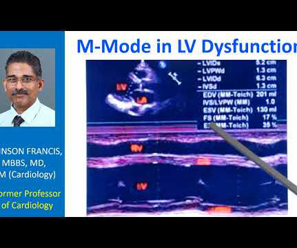
Transcript of the video: This is a still image of M-Mode Echocardiogram. Right ventricular outflow tract, left ventricle, left atrium, aorta, aorticvalve, mitral valve. M-Mode is Time-Motion Mode. The horizontal axis is time. Vertical axis is distance from the transducer. This is the parasternal long axis view.
You can see the reverse flow into the left ventricle from the aorta, with the aorticvalve in closed position. Beacuse, as the aorticvalve is closed, and the regurgitant jet is coming in, and left ventricular diastolic pressures are lower than the aortic pressures. Here it is the AR jet.
Transcript of the video: This is a still image from a colour Doppler echocardiogram, obtained from the apical five chamber view. Here, this is the forward flow through the mitral valve in diastole in red. This is reverse flow from the aorticvalve, that is aortic regurgitation jet. That also occurs in diastole.
Similarly, for echocardiogram, what we would do usually is, first we do a clinical history evaluation, then physical examination, and after that only we proceed with echocardiography in our routine work. You can see the two dimensional sector imaging from an echocardiogram and I have marked out the aorta.
Echocardiogram in parasternal long axis view shows dilated left ventricle, left atrium, aorta and a small portion of the right ventricle, which is usually the outflow region. Mitral valve leaflets seen in open position between the left ventricle and left atrium are thickened. Aorticvalve is seen as grossly thickened and calcified.
The image shown here is an animated 2 dimensional echocardiogram. This one is an older mode known as time-motion mode or M-Mode echocardiogram. An annotated video of normal colour Doppler echocardiogram illustrating various views and an M-Mode tracing is shown here. Exact position and angulation will vary between individuals.
We organize all of the trending information in your field so you don't have to. Join thousands of users and stay up to date on the latest articles your peers are reading.
You know about us, now we want to get to know you!
Let's personalize your content
Let's get even more personalized
We recognize your account from another site in our network, please click 'Send Email' below to continue with verifying your account and setting a password.


Let's personalize your content