This site uses cookies to improve your experience. To help us insure we adhere to various privacy regulations, please select your country/region of residence. If you do not select a country, we will assume you are from the United States. Select your Cookie Settings or view our Privacy Policy and Terms of Use.
Cookie Settings
Cookies and similar technologies are used on this website for proper function of the website, for tracking performance analytics and for marketing purposes. We and some of our third-party providers may use cookie data for various purposes. Please review the cookie settings below and choose your preference.
Used for the proper function of the website
Used for monitoring website traffic and interactions
Cookie Settings
Cookies and similar technologies are used on this website for proper function of the website, for tracking performance analytics and for marketing purposes. We and some of our third-party providers may use cookie data for various purposes. Please review the cookie settings below and choose your preference.
Strictly Necessary: Used for the proper function of the website
Performance/Analytics: Used for monitoring website traffic and interactions
The study, called IVUS-DCB, is the first randomized controlled trial to demonstrate the clinical benefits of using IVUS in angioplasty procedures for peripheral artery disease (PAD), a condition in which plaque builds up in arteries in the legs. Half were randomly assigned to receive IVUS plus angiography and half received angiography alone.
The goal of the ULTIMATE III trial was to evaluate intravascular ultrasound (IVUS)-guided drug-coated balloon angioplasty compared with angiography-guided drug-coated balloon angioplasty among patients with de novo coronary lesions.
The goal of the IVUS-DCB trial was to evaluate intravascular ultrasound (IVUS)-guided drug-coated balloon angioplasty compared with angiography-guided drug-coated balloon angioplasty among patients with femoropopliteal artery disease.
PTA+DCB, P Key findings include: · One-year primary patency (freedom from both clinically driven target lesion revascularization and duplex ultrasound-derived restenosis) did not differ between groups, despite the significant difference in baseline calcification. · DA+DCB versus 5.9% versus 21.1%, P =0.014). 3 · One-year rates of TLR (16.6%
In this week’s View, Dr. Eagle looks at the difference between quantitative coronary angiography versus intervascular ultrasound to guide PCI. He then discusses paclitaxel-coated balloon catheters vs uncoated balloon angioplasty for treating coronary in-stent restenosis.
Publication date: Available online 26 March 2025 Source: The American Journal of Cardiology Author(s): Jaeoh Lee, Ji Yong Jang, Chul-Min Ahn, Seung-Jun Lee, Sang-Hyup Lee, Yong-Joon Lee, Sung-Jin Hong, Jung-Sun Kim, Byeong-Keuk Kim, Myeong-Ki Hong, Yangsoo Jang, Tae-Hoon Kim, Ha-Wook Park, Jae-Hwan Lee, Jae-Hyeong Park, Su Hong Kim, Eui Im, Sang-ho (..)
Andreas Grüntzig was a German cardiologist best known for being the first to develop successful balloon angioplasty for expanding lumens of narrowed arteries. He also played a key role in developing intravascular ultrasound, as well as the U.S. standard-of-care for managing postpartum hemorrhage, the JADA System.
During the roundtable, participants highlighted the potential of IVUS in guiding revascularization procedures, such as angioplasty and stenting, to optimize outcomes for patients. It provides detailed information about the vessel wall, plaque composition, and blood flow characteristics, enabling more accurate diagnosis and treatment planning.
Interventional management involved performing percutaneous transluminal coronary angioplasty using balloon dilation on both the left main coronary artery and its ostium. Intravascular ultrasound confirmed successful dilation of the coronary ostium.
We aimed ultrasound-guided punctures in the proximal two-thirds of axillary arteries with diameters ≥2 mm to insert 7 cm/4 Fr short introducers. Overall, 27/36 procedures were interventional, including 6 aortic valvuloplasties, 6 balloon angioplasties, and 15 stenting procedures. We administrated intra-arterial verapamil (1.25 mg)
ET Main Tent (Hall B1) Coronary Sinus Reducer for the Treatment of Refractory Angina: A Randomised, Placebo-controlled Trial (ORBITA-COSMIC) Transcatheter Aortic Valve Implantation Versus Surgical Aortic Valve Replacement in Patients at Low to Intermediate Risk: One Year Outcomes of the Randomized DEDICATE-DZHK6 Trial Effect of Alcohol-mediated Renal (..)
After guidewire crossing, balloon angioplasty was performed, and a drug-eluting stent was deployed. An intravascular ultrasound was also performed, which was negative for vessel dissection. The left circumflex had 80% proximal stenosis with minimal luminal irregularities in the mid to distal portion.
Subjects were excluded if balloon angioplasty was performed prior to/within 2 days of the index procedure. Transcranial doppler (TCD) ultrasound was measured pre-IAT (D0) and at D+1 and D+2 post-IAT. All subjects received 10 mg IA milrinone, a non-catecholamine phosphodiesterase inhibitor drug, per MCA.
Bedside ultrasound with no apparent wall motion abnormalities, no pericardial effusion, no right heart strain. A comparison of electrocardiographic changes during reperfusion of acute myocardial infarction by thrombolysis or percutaneous transluminal coronary angioplasty. Aorta briefly viewed, appears normal caliber and diameter.
A lower extremity arterial ultrasound revealed elevated velocities in the right proximal superficial femoral artery. Based on these results, Dormu performed a percutaneous transluminal balloon angioplasty and a mechanical atherectomy and stenting of the right superficial femoral artery and stenting of the right superficial femoral artery.
Reports can be prepared for ultrasound, angioplasty, and TEE with the use of robust analysis. CONCLUSION The Cath lab technology for cardiologists reports tools that can prepare lengthy reports in a few minutes. After all, it has a plethora of pre-designed templates to help speed many quality reports in no time.
Discussing further, Catheterization Laboratory, also called Cath Lab, is a medical examination room where angiogram, angioplasty, ablation, and implantation of pacemaker are carried out. Ultrasound, TEE, and IVUS play an influential role in dropping the usage of angiographic imaging. Building Smart Cath Labs is highly related to it.
Spontaneous coronary artery dissection Dissection of a coronary artery may occur in the context of atherosclerosis, or be iatrogennic during angiography or angioplasty. Often, intravascular ultrasound or intravascular optical coherence tomography is requeried to make the diagnosis. This case occurred 10+ years ago.
His ED cardiac ultrasound (which is not at all ideal for detecting wall motion abnormalities, and is also very operator dependent for this finding) was significant for depressed global EF. The lesion was intervened on with balloon angioplasty and had subsequent TIMI 3 flow. The patient's initial troponin I was 2.0
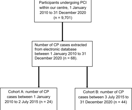
Factors associated with perforation include CTO or type C lesions and use of IVUS, cutting balloon angioplasty or hydrophilic wires. Conclusion The incidence of CP is increasing as more complex PCI is performed.
We organize all of the trending information in your field so you don't have to. Join thousands of users and stay up to date on the latest articles your peers are reading.
You know about us, now we want to get to know you!
Let's personalize your content
Let's get even more personalized
We recognize your account from another site in our network, please click 'Send Email' below to continue with verifying your account and setting a password.


Let's personalize your content