This site uses cookies to improve your experience. To help us insure we adhere to various privacy regulations, please select your country/region of residence. If you do not select a country, we will assume you are from the United States. Select your Cookie Settings or view our Privacy Policy and Terms of Use.
Cookie Settings
Cookies and similar technologies are used on this website for proper function of the website, for tracking performance analytics and for marketing purposes. We and some of our third-party providers may use cookie data for various purposes. Please review the cookie settings below and choose your preference.
Used for the proper function of the website
Used for monitoring website traffic and interactions
Cookie Settings
Cookies and similar technologies are used on this website for proper function of the website, for tracking performance analytics and for marketing purposes. We and some of our third-party providers may use cookie data for various purposes. Please review the cookie settings below and choose your preference.
Strictly Necessary: Used for the proper function of the website
Performance/Analytics: Used for monitoring website traffic and interactions
ABSTRACT Introduction Pulmonary vein (PV) restenosis develops with reported incidence rates of up to 50%. Balloon angioplasty seems to be the widely preferred treatment of choice. The stenosis was treated with a stent. The stenosis was treated with a stent. He has since remained asymptomatic over 3 years of follow-up.
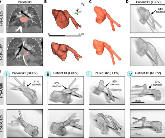
IntroductionPrimary pulmonary vein stenosis (PVS) is a rare congenital heart disease that proves to be a clinical challenge due to the rapidly progressive disease course and high rates of treatment complications. These 3D reconstructions were 3D printed using a clear resin ink and used in a benchtop experimental setup.
Our Interventional Cardiology Fellowship Program offers a unique opportunity for fellows to immerse themselves in high-volume centers and experience complex procedures such as angioplasties, stent placements, and Chronic Total Occlusion (CTO) interventions.
The combined and matched subgroups were pulmonary artery stenting (Stent PUL ), aorta angioplasty (Plasty AO ), pulmonary artery angioplasty (Plasty PUL ), or a combination of the latter two (Plasty). Three senior interventionists evaluated the relevance of MMIF 2D−3D (5-point Likert scale).
No signs for aortic dissection or pulmonary embolus. --"Results were discussed with the ordering physician. INTERVENTION * Successful angioplasty and stenting (drug eluting) of the mid LAD * Successful angioplasty of the ostial 1st diagonal Learning points: 1. A CT Coronary angiogram was ordered. CAD-RADS category 1. --No
Here is another proven left main occlusion in a young woman who presented with sudden pulmonary edema, had this ECG recorded, then arrested and was resuscitated after 30 minutes of CPR: This has sinus tachycardia with RBBB and LAFB, and STE in V2-V6 as well as I, aVL This pattern could just as easily be seen in LAD occlusion.
Once the patient reaches the hospital, the doctors will attempt to remove the clot using either a potent clot buster medicine [thrombolytic medicines] or a surgery known as primary angioplasty. Although both techniques have advantages and limitations, primary angioplasty is the chosen therapy in most cases.
We organize all of the trending information in your field so you don't have to. Join thousands of users and stay up to date on the latest articles your peers are reading.
You know about us, now we want to get to know you!
Let's personalize your content
Let's get even more personalized
We recognize your account from another site in our network, please click 'Send Email' below to continue with verifying your account and setting a password.


Let's personalize your content