
Post-stent chest pain, revisited
Heart Sisters
JULY 28, 2024
Heart patients with persistent or recurrent post-stent chest pain present “an unmet clinical need”, according to the European Journal of Cardiology.

Heart Sisters
JULY 28, 2024
Heart patients with persistent or recurrent post-stent chest pain present “an unmet clinical need”, according to the European Journal of Cardiology.

Dr. Smith's ECG Blog
JANUARY 31, 2020
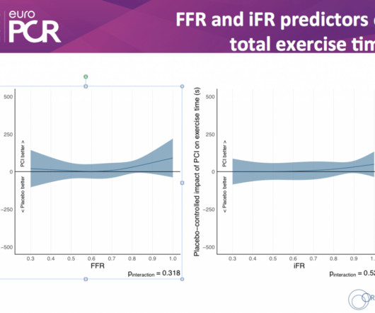
A middle-aged man complained of 15 minutes of classic angina that resolved upon arrival to the ED. Angiogram: Widely patent RCA and LAD stents. Therefore, no stent was placed. (No It is proven better than angiography alone in stable angina , and also has been shown to improve decisions on stenting non-culprit lesions in ACS.

Dr. Prateek Bhatnagar
MAY 11, 2022
A 55 years old diabetic male patient who had 12 stents in his heart underwent a successful beating heart bypass surgery under Dr. Prateek Bhatnagar, Director Cardiac Surgery. The patient was suffering with angina (chest pain) since 2002. He received these 12 stents on 5 different occasions at 5 different hospitals of the twin cities.

Dr. Prateek Bhatnagar
SEPTEMBER 6, 2023
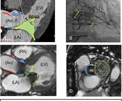
Prateek Bhatnagar Director Cardiac Surgery, on a 50 years man after stents placed in his left main coronary artery at Delhi just 3 months back, had blocked. Mr. Hemant, a resident of Delhi NCR, had developed chest pain (angina). He subsequently underwent stenting procedure in left main coronary artery.

Dr. Smith's ECG Blog
JANUARY 5, 2024
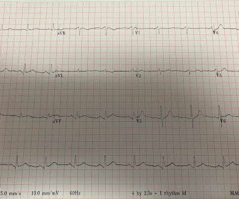
Thus, the patient does not (yet) get a formal diagnosis of MI and must be called unstable angina unless further troponins return above the 99th percentile. On the basis of unresolved angina, cardiology decided to perform rescue PCI. RCA and PDA before and after, arrows indicating stented regions. Repeat ECG is shown below.

Dr. Smith's ECG Blog
DECEMBER 19, 2023
The commonest causes of MINOCA include: atherosclerotic causes such as plaque rupture or erosion with spontaneous thrombolysis, and non-atherosclerotic causes such as coronary vasospasm (sometimes called variant angina or Prinzmetal's angina), coronary embolism or thrombosis, possibly microvascular dysfunction.

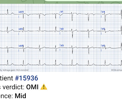
Dr. Smith's ECG Blog
APRIL 22, 2024
Patient is pain free and clearly has Wellens' syndrome: 1) pain free episode following an episode of angina, typical Pattern A (biphasic, terminal T-wave inversion with an initial upsloping ST Segment) findings, preserved R-waves. Angiography : --Culprit for the patient's unstable angina/Wellen syndrome is a ruptured plaque in the mid LAD. --As

Expert insights. Personalized for you.


Let's personalize your content