This site uses cookies to improve your experience. To help us insure we adhere to various privacy regulations, please select your country/region of residence. If you do not select a country, we will assume you are from the United States. Select your Cookie Settings or view our Privacy Policy and Terms of Use.
Cookie Settings
Cookies and similar technologies are used on this website for proper function of the website, for tracking performance analytics and for marketing purposes. We and some of our third-party providers may use cookie data for various purposes. Please review the cookie settings below and choose your preference.
Used for the proper function of the website
Used for monitoring website traffic and interactions
Cookie Settings
Cookies and similar technologies are used on this website for proper function of the website, for tracking performance analytics and for marketing purposes. We and some of our third-party providers may use cookie data for various purposes. Please review the cookie settings below and choose your preference.
Strictly Necessary: Used for the proper function of the website
Performance/Analytics: Used for monitoring website traffic and interactions
Coronary artery spasm (CAS), or Prinzmetal angina, is a recognised cause of myocardial ischaemia in non-obstructed coronary arteries which typically presents with anginal chest pain. This case report describes an atypical presentation of CAS in a 68-year-old white British male with cardiovascular risk factors.
Ischemia with no obstructive coronary arteries (INOCA) is an increasingly recognized condition in patients presenting with angina and positive stress tests but without significant coronary artery stenosis.
However, there is a notable absence of data regarding patients with short-term myocardial ischemia, such as those experiencing unstable angina (UA). In this report, we evaluated a 49-year-old male with UA and severe stenosis in multiple coronary arteries using 68 Ga-FAPI-04 PET/CT.
Likelihood of truth : High The flamboyant genius of Andreas Roland Gruntzig, from Zurich gifted us the path-breaking treatment modality for coronary stenosis five decades ago. No one has questioned the efficacy of PCI for true angina with a critical lesion. Transluminal dilatation of coronary-artery stenosis. Reference 1.
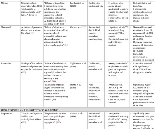
The goal of the DEFINE-FLAIR trial was to evaluate if functional lesion assessment by instantaneous wave-free ratio (iFR) would be noninferior to fractional flow reserve (FFR) among patients with stable angina or acute coronary syndromes.
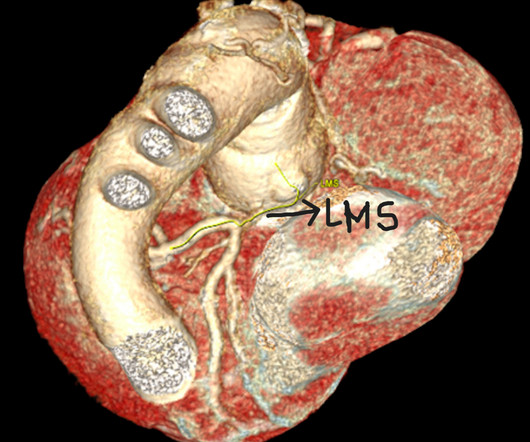
Such a pattern is consistent with significant left main coronary artery stenosis. Angiography done after initial stabilization showed severe stenosis of distal left main coronary artery. This patient had reported with recent onset angina. ST segment elevation is noted in aVR. ST elevation was 2 mm in aVR and 1 mm in V1.
Objectives There are few reports regarding the prognosis in patients with obstructive coronary artery disease (OCAD) and vasospastic angina (VSA). We defined positive epicardial spasm as ≥90% transient stenosis and usual chest symptoms or ischemic ECG changes. An obstructed coronary artery was defined as ≥50% luminal narrowing.
Six months following modified Bentall procedure a patient presented with angina and acute ST depression. CT coronary angiogram revealed severe narrowing of the left main coronary artery. Minimal invasive off p.
Since the pathologist does not know the original cross-sectional area of the artery or the amount of compensatory enlargement of the artery from evaluation of a single cross section of the artery at a site of stenosis, the degree of luminal narrowing of that segment cannot be determined. These are typical findings at sites of plaque rupture.
A middle-aged man complained of 15 minutes of classic angina that resolved upon arrival to the ED. Culprit Lesion: Angiographically indeterminate 50% stenosis in the proximal OM2 was assessed further with instantaneous wave free ratio (iFR) of 0.96, which is normal (see below for description of iFR*).
Patient is pain free and clearly has Wellens' syndrome: 1) pain free episode following an episode of angina, typical Pattern A (biphasic, terminal T-wave inversion with an initial upsloping ST Segment) findings, preserved R-waves. Angiography : --Culprit for the patient's unstable angina/Wellen syndrome is a ruptured plaque in the mid LAD. --As
However, revascularisation of coronary stenosis, which induces myocardial ischaemia, has demonstrated better outcome than OMT alone. Patients with diffuse disease showed a twofold risk of residual angina after percutaneous coronary intervention (PCI) than those with focal disease.
Given the consistency of the clinical profile with typical angina, associated risk factors, and abnormal ECG findings, a cardiology consult was promptly requested. Aortic Stenosis f. Left Main stenosis (not thrombosed) c. Aortic Stenosis [No Hx syncope, and no systolic murmur] f. Left Main stenosis (not thrombosed) c.
Thus, the patient does not (yet) get a formal diagnosis of MI and must be called unstable angina unless further troponins return above the 99th percentile. On the basis of unresolved angina, cardiology decided to perform rescue PCI. The LAD has diffuse disease with a few areas of moderate stenosis but no flow-limiting lesions.
Background Aortic stenosis is a life-limiting condition for which transcatheter aortic valve implantation (TAVI) is an established therapy. Subsidiary outcomes included patient angina and breathlessness scores. CAD at the time of TAVI also did not alter breathlessness or angina scores before/after TAVI (p>0.05).
link] A 62 year old man with a history of hypertension, type 2 diabetes mellitus, and carotid artery stenosis called 911 at 9:30 in the morning with complaint of chest pain. This is written by Willy Frick, an amazing cardiology fellow in St. He described it as "10/10" intensity, radiating across his chest from right to left.
The primary end point was minimal lumen area at 6-month follow-up.RESULTS:Following Magmaris BRS implantation, minimal lumen area (6.41.6 versus 6.31.5 mm2;P=0.65), mean scaffold area (7.81.5 versus 7.51.7 mm2;P=0.37), and mean lumen area (8.01.6 versus 7.72.1
This appears to be a classic Wellens' ECG, Pattern A, with terminal T-wave inversion in V2-V4, preserved R-waves, and it appears to be Wellens' syndrome, as it occurred after resolution of typical angina pain. When this happens, troponins are negative, there is no wall motion abnormality, and it is true unstable angina.
If it is angina, lowering the BP with IV Nitroglycerine may completely alleviate the pain and the (unseen) ECG ischemia. Or is it a very tight stenosis that does not allow enough flow to perfuse myocardium that has a high oxygen demand from severely elevated BP? The ST depressions in I and aVL have resolved.
Methods We retrospectively enrolled 237 patients who underwent elective coronary angiography (CAG) for stable angina or positive stress test results >1 year after CABG. The SVGD was defined as presence of at least 50% stenosis in at least 1 SVG. The patients were divided into two groups; SVGD (+) patients and SVGD (−) patients.
SMART 4 ( NCT04722250 ) studied patients with severe aortic stenosis and a small aortic annulus who underwent transcatheter aortic valve replacement (TAVR). The primary endpoint consisted of a composite of all-cause mortality, MI, stroke, coronary revascularization, or hospitalization for unstable angina. vs. 6.2%) and stroke (2.9%
When there is extremely brief ischemia, as in this case , or this case , it may entirely reverse, especially in unstable angina (negative troponins). Angiographic and clinical characteristics of patients with unstable angina showing an ECG pattern indicating critical narrowing of the proximal LAD coronary artery. Lessons: 1.
Notably, the LAD had multiple aneurysmal segments and areas of eccentric stenosis upto 90%.Multislice However some patients can develop heart failure, angina, and arrhythmia due to significant intracardiac shunt or coronary steal phenomenon. CCF can be congenital or acquired and has many variations. It is often clinically silent.
Patients with dextrocardia present a diagnostic challenge, particularly in the context of acute coronary syndrome.Case Presentation:A 49-year-old male with a medical history of dextrocardia, hypothyroidism, dyslipidemia and hypertension was referred to a cardiologist by his primary physician due to a 3-week history of unstable angina.
Takotsubo is a sudden event, not one with crescendo angina. 1-4 Surprisingly, serial angiographic studies have revealed that the plaque at the site of the culprit lesion of a future acute myocardial infarction often does not cause stenosis that, as seen on the antecedent angiogram, is sufficiently severe to limit flow.
Typical angina was defined as a symptom complex that includes substernal chest pressure or pain that was made worse with exertion/emotional stress, and relieved by rest or nitroglycerin. Atypical angina is classified as having any two of the three symptoms, and non-anginal pain any one of the three symptoms.
24: Joint American College of Cardiology/Journal of the American College of Cardiology Late-Breaking Clinical Trials (Session 402) Saturday, April 6 9:30 – 10:30 a.m.
Serial EKG is very high yield diagnostic test in patients with stuttering angina. If a patient’s presentation is suggestive of stress cardiomyopathy, the onus is on the treating clinician to rule out other causes including OMI. EKG 3 is diagnostic for developing re-occlusion, and EKG 4 proves that the nitrates relieved the ischemia. =
The patient had a critical LAD stenosis. Tight proximal LAD stenosis explains STE in precordial leads and I and aVL. I did not include the prehospital because it is identical to the first ED ECG: Self explanatory, no? All troponins were u ndetectable (less than 0.04 Flow had spontaneously been restored, perhaps aided by nitroglycerin.
LAD plaque with 0-25 percent stenosis. The LAD has moderate 40% ostial-proximal LAD stenosis and severe 90% mid LAD stenosis involving first diagonal branch. --The If trops are negative and there is <50% stenosis, then the patient is safe for discharge, even if the HEART score or EDACS score are elevated.
Beware crescendo angina in patient with known CAD ST Elevation in aVR Case 7. baseline LVH, demand ischemia secondary to respiratory failure, aortic stenosis, hemorrhagic shock). found that only 23% of patients with the aVR STE pattern had any LM disease (fewer if defined as ≥ 50% stenosis). This was a 100% acute LM occlusion.
Characteristic electrocardiographic pattern indicating a critical stenosis high in left anterior descending coronary artery in patients admitted because of impending myocardial infarction. Am Heart J. 1982 Apr;103(4 Pt 2):730-6. de Zwaan C et al.
Here are the images from the cardiac cath: Mid to distal-LAD in-stent stenosis with 100% occlusion and TIMI flow 0 LAD post-DES placement with TIMI 3 flow The amount of territory supplied by this vessel becomes obvious here (and goes on for a few more frames below this still).
We evaluated the primary outcome (cardiovascular death, myocardial infarction, or hospitalization for unstable angina, heart failure, or resuscitated cardiac arrest) and other end points, by sex, in 1168 (22.6%) women and 4011 (77.4%) men. of invasive‐assigned men, and no ≥50% stenosis in 12.3% of invasive‐assigned men;P<0.001).
Angiograms were examined for luminal stenosis in each segment of the SYNTAX coronary model. Cumulative plaque burden was quantified using the Gensini score, which incorporated both the number of diseased coronary segments and stenosis severity. –11.0)), vasospastic angina (VSA) (4.5 (2.0–10.0)),
He was diagnosed with an unstable angina, and coronary angiography showed near-total in-stent occlusion of the previously placed stent protruding into the aorta. Coronary angiography revealed tight stenosis at the ostial left anterior descending artery with a previous stent deployed from the left main to the circumflex artery.
Angiogram: Severe two-vessel coronary artery disease of a left dominant system including 70 to 80% stenosis involving the distal left main/bifurcation. The estimated left ventricular ejection fraction is 64%. There is no left ventricular wall motion abnormality identified. The ECG shows inferior ischemia.
We organize all of the trending information in your field so you don't have to. Join thousands of users and stay up to date on the latest articles your peers are reading.
You know about us, now we want to get to know you!
Let's personalize your content
Let's get even more personalized
We recognize your account from another site in our network, please click 'Send Email' below to continue with verifying your account and setting a password.


Let's personalize your content