This site uses cookies to improve your experience. To help us insure we adhere to various privacy regulations, please select your country/region of residence. If you do not select a country, we will assume you are from the United States. Select your Cookie Settings or view our Privacy Policy and Terms of Use.
Cookie Settings
Cookies and similar technologies are used on this website for proper function of the website, for tracking performance analytics and for marketing purposes. We and some of our third-party providers may use cookie data for various purposes. Please review the cookie settings below and choose your preference.
Used for the proper function of the website
Used for monitoring website traffic and interactions
Cookie Settings
Cookies and similar technologies are used on this website for proper function of the website, for tracking performance analytics and for marketing purposes. We and some of our third-party providers may use cookie data for various purposes. Please review the cookie settings below and choose your preference.
Strictly Necessary: Used for the proper function of the website
Performance/Analytics: Used for monitoring website traffic and interactions
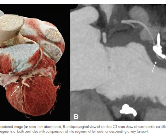
Inflammatory pericarditis can occur in differential fashion. For example, the most common chronic pericarditis tuberculosis affects the fibrinous layer. Post MI pericarditis involves the epicardium. There has been many reports of patients with angina in CP (Ref 1). Angina caused by calcific constrictive pericarditis.
Pericarditis refers to inflammation of the pericardium The pericardium is a sac within which the heart sits. Acute inflammation of this sac is known as acute pericarditis. About 5% of patients who present to A+E with chest pain which is not deemed to be a heart attack or angina are ultimately diagnosed with pericarditis.
06:44 - T-waves in V2 are smaller now - Overall resolution of prior findings (which qualifies as a dynamic change) The initial note by the cardiologist states that the presentation is more consistent with pericarditis. Remember, pericarditis is the thing you say and write down when youre actively trying to miss an OMI.
This is a value typical for a large subacute MI, n ormal value 48 hours after myocardial infarction is associated with Post-Infarction Regional Pericarditis ( PIRP ). As already mentioned, this patient could have post-infarction regional pericarditis from a large completed MI. Sinus tachycardia has many potential causes. Hammill SC.
The typical pain of cardiac origin is a central chest pain which occurs on walking or other forms of exercise, known as effort angina. Effort angina is commonly due to significant obstruction to a blood vessel (coronary artery) supplying a part of the heart muscle. Pain is likely to be more if you are walking after a heavy meal.
This rules out pericarditis, which essentially never has reciprocal ST depression. This is not pericarditis because: a. Pericarditis does not have reciprocal depression. ST elevation of pericarditis is maximal in leads II and V5, V6. Pericarditis does not have hyperacute T-waves.
A middle-aged woman had intermittent angina for 48 hours, then onset of constant, crushing chest pain for 1.5 More likely, the patient had crescendo angina, with REVERSIBLE ischemia for 48 hours that only became potentially irreversible (STEMI) at that point in time. Myocardial Rupture and Postinfarction Pericarditis.
OMI is generally of more acute onset, unless there is intermittent angina. There is also mild pericardial enhancement consistent with pericarditis. I learned more about the history: 30-something African American with 5-7days of sharp R-sided shoulder/scapula/chest discomfort, presented with sinus tachycardia.
That is usually with angina and ventricular strain patterns. PR segment elevation and depression can occur in atrial infarction and pericarditis. So in anterior leads, for diagnosis of ST elevation myocardial infarction, V1, the cutoff is usually 2 mm, while 1 mm is enought in other leads. When there is ST depression, even 0.5
The patient might be having cardiac ischemia, but if he is, it is unstable angina or non-STEMI, or perhaps he has not YET pseudonormalized, so serial ECGs may be important. Differential of peri-infarct pericardial fluid The differential includes 1) pericarditis with effusion or 2) hemopericardium. Myocardial rupture is not uncommon.
The exception is with postinfarction pericarditis , in which a completed transmural infarct results in inflammation of the subepicardial myocardium and STE in the distribution of the infarct, and which results in increased STE and large upright T-waves. These findings together are more commonly seen with pericarditis.
We organize all of the trending information in your field so you don't have to. Join thousands of users and stay up to date on the latest articles your peers are reading.
You know about us, now we want to get to know you!
Let's personalize your content
Let's get even more personalized
We recognize your account from another site in our network, please click 'Send Email' below to continue with verifying your account and setting a password.


Let's personalize your content