This site uses cookies to improve your experience. To help us insure we adhere to various privacy regulations, please select your country/region of residence. If you do not select a country, we will assume you are from the United States. Select your Cookie Settings or view our Privacy Policy and Terms of Use.
Cookie Settings
Cookies and similar technologies are used on this website for proper function of the website, for tracking performance analytics and for marketing purposes. We and some of our third-party providers may use cookie data for various purposes. Please review the cookie settings below and choose your preference.
Used for the proper function of the website
Used for monitoring website traffic and interactions
Cookie Settings
Cookies and similar technologies are used on this website for proper function of the website, for tracking performance analytics and for marketing purposes. We and some of our third-party providers may use cookie data for various purposes. Please review the cookie settings below and choose your preference.
Strictly Necessary: Used for the proper function of the website
Performance/Analytics: Used for monitoring website traffic and interactions
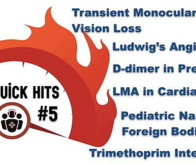
The post EM Quick Hits 5 Ludwig’s Angina, Transient Monocular Vision Loss, D-dimer for PE Workup in Pregnancy, Pediatric Nasal Foreign Bodies, Trimethoprim Drug Interactions, Airway Management in Cardiac Arrest appeared first on Emergency Medicine Cases.
The disease can cause a variety of symptoms, including heart failure, arrhythmias, peripheral embolism, dysautonomia, angina with normal coronary arteries, and others. The aim of this thematic collection is to gather high-quality articles that contribute to the advancement of knowledge about cardiac amyloidosis.
The commonest causes of MINOCA include: atherosclerotic causes such as plaque rupture or erosion with spontaneous thrombolysis, and non-atherosclerotic causes such as coronary vasospasm (sometimes called variant angina or Prinzmetal's angina), coronary embolism or thrombosis, possibly microvascular dysfunction.
Reduction in admissions for stroke recurrence or related to stroke, (heart attack, angina, peripheral embolism, etc.): Stroke, 30days: -100%; Related to stroke, (365d: -47,7%; 30d: -57,0%). Reduction in cardiovascular admissions ( 30d: -100%; 365d: -31,4%).
The ECG is rather classic for pulmonary embolism, and indeed this was a large acute PE. This is NOT Wellens because the T-wave inversion is DURING the pain (not after -- Wellens' is a syndrome of TWI after an episode of angina is resolved). this is highly suggestive of pulmonary embolism. This is a classic S1Q3T3. Kosuge et al.
ET Main Tent (Hall B1) A Selective Aldose Reductase Inhibitor (at-001) For the Treatment of Diabetic Cardiomyopathy: Primary Results of the Phase 3 Randomized Controlled ARISE-HF Study Efficacy and Safety of Ninerafaxstat, a Novel Cardiac Mitotrope, in Patients with Symptomatic Nonobstructive Hypertrophic Cardiomyopathy: Results of IMPROVE-HCM Topical (..)
Coronary artery embolism – In this scenario, a blood clot which forms elsewhere goes down a coronary artery, causing a blockage but by the time we do the angiogram, the clot has dissipated and we see unobstructed vessels. Here is a video I have done on this subject.
A middle-aged woman had intermittent angina for 48 hours, then onset of constant, crushing chest pain for 1.5 Both of these are very suggestive of " No-Reflow ," or poor microvascular reperfusion due to downstream embolization of microscopic platelet-fibrin aggregates. hours when she called 911.
If there are T-wave inversions and elevated trops in the context of persistent pain, think of other pathologies such as pulmonary embolism. Wellens' syndrome will almost always develop elevated troponins. Wellens' syndrome is a post-OMI syndrome after spontaneous reperfusion. It is likely that the artery will re-occlude.
Angina is another common symptom due the hypertrophy which causes a coronary supply demand mismatch Symptoms of HCM include syncope/near syncope, which could be precipitated by exertion, hypovolemia, rapid standing, Valsalva manoeuvre, diuretics, vasodilators or arrhythmia. Doppler echo showing LVOT gradient in HCM.
Takotsubo is a sudden event, not one with crescendo angina. 9 This dissociation between the degree of stenosis and the propensity to provoke an acute coronary syndrome helps to explain why myocardial infarction often occurs without being heralded by the demand-induced symptoms of angina that would result from a high-grade stenosis.
Today's patient is a 50-something year old man who presented with increasingly severe exertional angina, for which his CP ( C hest P ain ) was just resolving at the time his initial ( triage ) ECG was recorded ( TOP tracing in Figure-1 ). By itself seeing this ECG pattern does not necessarily mean that the patient has a pulmonary embolism.
I have said before, treating angina with morphine and continuing non-emergent management is like taking the batteries out of an actively alarming smoke detector during a house fire and going back to sleep. And it is definitely possible that a more proximal LCx lesion ruptured and produced distal embolism.
We organize all of the trending information in your field so you don't have to. Join thousands of users and stay up to date on the latest articles your peers are reading.
You know about us, now we want to get to know you!
Let's personalize your content
Let's get even more personalized
We recognize your account from another site in our network, please click 'Send Email' below to continue with verifying your account and setting a password.


Let's personalize your content