This site uses cookies to improve your experience. To help us insure we adhere to various privacy regulations, please select your country/region of residence. If you do not select a country, we will assume you are from the United States. Select your Cookie Settings or view our Privacy Policy and Terms of Use.
Cookie Settings
Cookies and similar technologies are used on this website for proper function of the website, for tracking performance analytics and for marketing purposes. We and some of our third-party providers may use cookie data for various purposes. Please review the cookie settings below and choose your preference.
Used for the proper function of the website
Used for monitoring website traffic and interactions
Cookie Settings
Cookies and similar technologies are used on this website for proper function of the website, for tracking performance analytics and for marketing purposes. We and some of our third-party providers may use cookie data for various purposes. Please review the cookie settings below and choose your preference.
Strictly Necessary: Used for the proper function of the website
Performance/Analytics: Used for monitoring website traffic and interactions
Coronary aneurysm, a dilated segment of the coronary artery, is a rare condition with a prevalence ranging from 0.02% to 0.2%. According to the current literature, reports of large aneurysms in the left main artery are extremely rare. Due to the high surgical risk, conservative treatment was chosen.
Ruptured spinal artery aneurysms, <1% of SAH, pose challenges in understanding and management. In contrast to intracranial aneurysms, they often manifest with symptoms like back pain and possibly myelopathy. Our study presents five cases of spinal SAH from posterior spinal artery aneurysms.
IntroductionThe use of detachable coils for endovascular embolization of cerebral aneurysms has become a safe and effective alternative to direct surgical clipping in patients with ruptured aneurysmal subarachnoid hemorrhage. Immediate complete occlusion and occlusion with residual neck was achieved in 66.7%
A 9-day-old male neonate was found to have a systolic murmur during a routine follow-up for skin jaundice. Imaging revealed a large mass at the bifurcation of the main pulmonary artery, causing significant bil.
Previous medical interventions included a spectrum of procedures, including catheter-directed thrombectomy for popliteal artery aneurysms with thrombosis, vascular bypass grafting for cerebral-anterior communicating artery aneurysms and arch replacement and stent implantation for aortic dissecting aneurysms.
Old ‘NSTEMI’ A history of coronary artery disease and a stent to the same territory further increases pre-test likelihood of acute coronary occlusion, including in-stent thrombosis. Smith : Old inferior MI with persistent ST Elevation ("inferior aneurysm") has well-formed Q-waves. Does this change your interpretation?
IntroductionIndications for flow diversion for the treatment of cerebral aneurysms have increased remarkably in recent years.1 1 This has been particularly useful for aneurysms that are difficult to treat via endosaccular or open approaches, such as pseudoaneurysms.2
Coronary angiography revealed a tortuous and extremely aneurysmal RCA, as well as multivessel coronary artery disease (mvCAD) involving LAD, D1, LCx, OM1. Notably, the LAD had multiple aneurysmal segments and areas of eccentric stenosis upto 90%.Multislice
IntroductionSubarachnoid Hemorrhage (SAH) resulting from the spontaneous rupture of an aneurysm is a rare and highly debilitating condition. Despite its severity, patients with aneurysmal SAH remain understudied, particularly concerning the evaluation of the incidence and consequences of subsequent acute kidney injury (AKI).
Repeat CT angio chest (not CT coronary, unclear what protocol) showed possible LAD aneurysm and thrombus. Finally, coronary angiography was performed (at least 5 days after presentation) which confirmed LAD aneurysm with large thrombus burden, TIMI 0 flow, thrombectomy performed. No further cath details available.
1,2 ASCVD causes or contributes to conditions that include coronary artery disease (CAD), cerebrovascular disease, and peripheral vascular disease (inclusive of aortic aneurysm).3 Atherosclerotic cardiovascular disease (ASCVD), caused by plaque buildup in arterial walls, is one of the leading causes of disability and death worldwide.1,2
The patient's heart had significant recovery: Echo : Estimated LVEF 32%, apical wall motion abnormality with diastolic distortion (LV aneurysm), suggestive of old MI. Coronary thrombosis or embolism can result in MINOCA, either with or without a hypercoagulable state. It was uncertain whether this represented: 1.
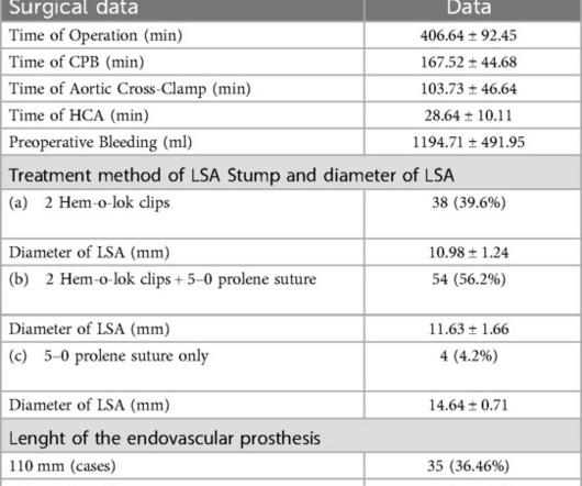
BackgroundFormation of local type aortic aneurysm years after surgical repair of coarctation (CoA) occurs in 10% of patients independent of the surgical technique and is a potentially life-threatening condition if left untreated with a high risk of aortic rupture.
Despite the widespread use of mini-invasive treatment methods in cardiac surgery, their use in post-infarction myocardial aneurysms of the left ventricle is not of frequent occurrence.
We organize all of the trending information in your field so you don't have to. Join thousands of users and stay up to date on the latest articles your peers are reading.
You know about us, now we want to get to know you!
Let's personalize your content
Let's get even more personalized
We recognize your account from another site in our network, please click 'Send Email' below to continue with verifying your account and setting a password.


Let's personalize your content