This site uses cookies to improve your experience. To help us insure we adhere to various privacy regulations, please select your country/region of residence. If you do not select a country, we will assume you are from the United States. Select your Cookie Settings or view our Privacy Policy and Terms of Use.
Cookie Settings
Cookies and similar technologies are used on this website for proper function of the website, for tracking performance analytics and for marketing purposes. We and some of our third-party providers may use cookie data for various purposes. Please review the cookie settings below and choose your preference.
Used for the proper function of the website
Used for monitoring website traffic and interactions
Cookie Settings
Cookies and similar technologies are used on this website for proper function of the website, for tracking performance analytics and for marketing purposes. We and some of our third-party providers may use cookie data for various purposes. Please review the cookie settings below and choose your preference.
Strictly Necessary: Used for the proper function of the website
Performance/Analytics: Used for monitoring website traffic and interactions
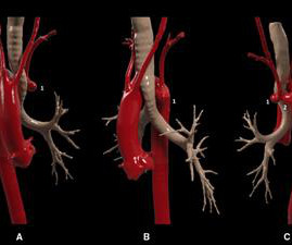
Imaging techniques and modeling technology allow a more personalized strategy for each patient. In this case, we present a symptomatic patient with a Kommerell's diverticulum and a left aberrant subclavian artery complicated by proximal stenosis and poststenotic aneurysm.
IntroductionThe accuracy of fenestrations in stent grafts for complex aortic aneurysms and dissections can be significantly improved using three-dimensional (3D)-printed phantoms. Standardization is enhanced by using artificial intelligence (AI) for image pre-processing before 3D printing.
Objective This study aims to review the application of deep learning techniques in the imaging diagnosis and treatment of aortic aneurysm (AA), focusing on screening, diagnosis, lesion segmentation, surgical assistance, and prognosis prediction. Furthermore, models were able to predict AA progression and patient prognosis with high accuracy.
IntroductionIndications for flow diversion for the treatment of cerebral aneurysms have increased remarkably in recent years.1 1 This has been particularly useful for aneurysms that are difficult to treat via endosaccular or open approaches, such as pseudoaneurysms.2
3D rotational angiography is not that old technique, but a novel technology which can create a volumetric data set of contrast-enhanced CT like images with excellent visualization of soft tissues and airway, with spatial resolution even better than multi-detector CT or cardiac magnetic resonance imaging [1].
Introduction:Although flow diverters (FD) are a long-established treatment-option for brain aneurysms, the evidence of data associated with therapy success is still based on relatively small heterogeneous studies. Median diameter and neck width of aneurysms were 7.2 91.7% (719) of aneurysms were saccular. mm (IQR, 5-11) and 6.5
Aortic Aneurysms : An aneurysm is an abnormal bulge in a blood vessel wall. Vasculitis : Inflammation of blood vessels that can lead to organ damage or an aneurysm. Angioplasty & Stenting: Opens blocked arteries to improve blood flow. This can lead to a transient ischemic attack (TIA) or stroke.
We organize all of the trending information in your field so you don't have to. Join thousands of users and stay up to date on the latest articles your peers are reading.
You know about us, now we want to get to know you!
Let's personalize your content
Let's get even more personalized
We recognize your account from another site in our network, please click 'Send Email' below to continue with verifying your account and setting a password.


Let's personalize your content