This site uses cookies to improve your experience. To help us insure we adhere to various privacy regulations, please select your country/region of residence. If you do not select a country, we will assume you are from the United States. Select your Cookie Settings or view our Privacy Policy and Terms of Use.
Cookie Settings
Cookies and similar technologies are used on this website for proper function of the website, for tracking performance analytics and for marketing purposes. We and some of our third-party providers may use cookie data for various purposes. Please review the cookie settings below and choose your preference.
Used for the proper function of the website
Used for monitoring website traffic and interactions
Cookie Settings
Cookies and similar technologies are used on this website for proper function of the website, for tracking performance analytics and for marketing purposes. We and some of our third-party providers may use cookie data for various purposes. Please review the cookie settings below and choose your preference.
Strictly Necessary: Used for the proper function of the website
Performance/Analytics: Used for monitoring website traffic and interactions
Following SAH, neutrophils cause vascular occlusion via neutrophil extracellular traps (NETs) and NETs have been identified as a therapeutic target to prevent delayed cerebral ischemia in mice (DCI) with SAH. The findings of this study suggest NETs factors may be useful biomarkers to predict which aneurysmal SAH patients are at-risk for DCI.
Kawasaki disease (KD), an acute self-limited febrile illness that primarily affects children <5 years old, is the leading cause of acquired heart disease in developed countries, with the potential of leading to coronary artery dilation and coronary artery aneurysms in 25% of untreated patients.
There were many comments that it was too late for thrombolytics or that this signified an LV aneurysm, not acute MI. See my formula for differentiating anterior LV aneurysm (that is to say, persistent ST elevation after old MI) from acute anterior STEMI. See my answer below. This is my response: "This is definitely acute or subacute.
ObjectiveSpinal cord ischemia due to damage or occlusion of the orifices of aortic segmental arteries (ASA) is a serious complication of open and endovascular aortic repair.
6 Ruptured intracerebral aneurysms are associated with higher morbidity and mortality than unruptured ones. This is secondary to delayed postoperative cerebral ischemia and infarction caused by vasospasm.7 This is secondary to delayed postoperative cerebral ischemia and infarction caused by vasospasm.7
BACKGROUNDThe optimal endovascular approach for acutely ruptured wide‐neck intracranial aneurysms remains uncertain, and the use of stent‐assisted coiling or flow diversion is controversial due to antiplatelet therapy requirements and potential risks. Stroke: Vascular and Interventional Neurology, Ahead of Print. versus BAC: 2.8%;P=
Approximately 30% of aneurysmal subarachnoid hemorrhage (aSAH) patients who survive the rupture develop delayed cerebral ischemia (DCI) 4 to 10 days following aSAH. Stroke, Volume 56, Issue Suppl_1 , Page AWP370-AWP370, February 1, 2025.
IntroductionThe optimal endovascular approach for wide‐neck intracranial aneurysms (IAs) during the acute phase of bleeding remains uncertain, and the use of stent‐assisted coiling or flow diversion is controversial due to antiplatelet therapy requirements and potential risks (1, 2).
Introduction:Delayed cerebral ischemia (DCI) is a leading cause of morbidity and mortality in aneurysmal subarachnoid hemorrhage (aSAH). There was no significant difference between the two arms with regards to admission characteristics, aneurysm location, treatment type, or total number of vasospasm treatments.
In this study, our hypothesis was that NETs cause vascular occlusion leading to delayed cerebral ischemia (DCI) and worse outcome after SAH. Aneurysmal SAH patients who developed DCI had elevated markers of NETs compared to non-DCI patients. Similar findings were observed for PAD4 inhibition.
Introduction:Elevated levels of Interleukin-6 (IL-6) levels in cerebrospinal fluid (CSF) have been correlated with delayed cerebral ischemia (DCI) after aneurysmal subarachnoid hemorrhage (aSAH). Stroke, Volume 55, Issue Suppl_1 , Page AWP152-AWP152, February 1, 2024.
Mechanism is thought to be due to sustained sympathetic stimulation, probably caused by dysfunction of insular cortex resulting in reversible neurogenic damage to the myocardium which could include contraction bands and subendocardial ischemia [2]. Serial measurements of cardiac enzymes were normal in that case. 2009 Nov;40(11):3478-84.
However, old MI w/aneurysm morphology (persistent ST-Elevation) can look just like this. While this may be change that is reciprocal to an Acute/Subacute Inferior STEMI, the problem is that LV aneurysm may also manifest with this reciprocal change. Old MI w/Aneurysm will show moderate ST Elevation, as seen here.
One very useful adjunct is ultrasound: Echo of his heart can distinguish aneurysm from acute MI by presence of diastolic dyskinesis, but it cannot distinguish demand ischemia from ACS. These must raise suspicion of old MI with persistent ST elevation. The patient was suffering from severe dehydration, possibly with sepsis.
Are you confident there is no ischemia? Primary VT , and the VT with tachycardia is causing ischemia with chest discomfort (supply-demand mismatch/type 2 MI)? Ischemia from ACS causing the chest discomfort, with VT another consequence (or coincidence)? These are all findings that can be expected with left ventricular aneurysm.
Normal RBBB, no evidence of ischemia. This may be permanent and may be associated with echocardiographic dyskinesis (aneurysm). LV aneurysm is common in completed, full thickness (transmural) MI, which is what we have here. Here is the patient's previous ECG (Figure 2): Previous ECG. R-waves of of normal height.
The old ECG has a Q-wave with persistent ST elevation in lead III, and some reciprocal ST depression (typical for aneurysm morphology). This is "Persistent ST elevation after previous MI" or "LV aneurysm morphology". LV aneurysm is very different for inferior vs. anterior MI. This is not pericarditis because: a.
All of this appears to be consistent with "No Reflow", or small vessel occlusion with persistent ischemia in spite of an open artery. This ECG is diagnostic of anterior LV aneurysm in the presence of RBBB. See more such cases of RBBB with LV aneurysm here. He was hospitalized for 16 days. ECG one month later: What do you think?
Of the 32 patients, 9(28.1%) had dissection with diagnostic angiograms, 6(18.8%) endovascular thrombectomy, 15(46.9%) aneurysm treatment, and 2(6.3%) angioplasty with or without stenting. Common comorbidities included hypertension (62.5%), smoking (56.3%), and hyperlipidemia (46.9%).
Methods:This was a retrospective observational study using data from the large multicenter international Stroke Thrombectomy and Aneurysm Registry (STAR). The primary outcome measure was successful recanalization defined as modified Thrombolysis in Cerebral Ischemia (mTICI) score of 2b or higher.
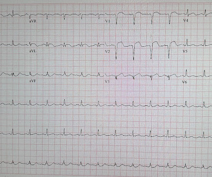
T-wave inversion in V2 is inconsistent with early repol, and is typical of posterior ischemia. In addition, there is ST depression, diagnostic of ischemia, in V3-V6. A coronary aneurysm was found. T-waves in inferior leads are hyperacute, out of proportion to those in early repol 5. mm of ST elevation in inferior leads.
I do not think this ECG is by itself diagnostic of OMI (full thickness, subepicardial ischemia ), b ut comparison to a previous might reveal this ECG as diagnostic of OMI. A CT was completed to rule out dissection, PE, or aneurysm, and this was unremarkable. mg/dL, K 3.5 Another 162mg ASA and heparin were given at this point.
The aim of this study is to develop and validate a stroke prediction tool for outcome in MT for AIS patients with low ASPECTS using data from an ongoing international multicenter registry, the Stroke Thrombectomy and Aneurysm Registry (STAR).Methods:236 90-day mRS 0-3 was observed in 0% of patients with a score of 0 or 1, 6.3%
Persistent ST elevation 3 days after a nearly transmural MI portends possible LV aneurysm. It is very unlikely to be LV aneurysm morphology when the ST elevation is so high and the T-Wave inversion is so deep. The patient continued to have ischemia after PCI, and in fact had an episode of polymorphic VT shortly after while in the ICU.
The only time you see this without ischemia is when there is an abnormal QRS, such as LVH, LBBB, LV aneurysm (old MI with persistent STE) or WPW." Here is the patient's troponin I profile: These were interpreted as due to demand ischemia, or type II MI. ng/mL is seldom a result of demand ischemia (type 2 MI).
My rule is that if any lead has a T/QRS ratio >0.36, then it is acute OMI; if <0.36, then either old MI (LV aneurysm) or subacute OMI. There is ST depression in V3 and V4. Always abnormal.
However, the ST segments in patients with LVH may show significant variation over time in the absence of ischemia. 3 Some have also suggested that the typically asymmetric T wave inversion (TWI) of LVH might be distinguished from the typically symmetric TWI of cardiac ischemia. Is LVH like left ventricular aneurysm?
Prior ECG available on file from 2 months before: We do not know the clinical events happening during this ECG, but there is borderline tachycardia, PVCs, and likely some evidence of subendocardial ischemia with small STDs maximal in V5-6/II, slight reciprocal STE in aVR. QS waves from V2-V5 consistent with LV aneurysm morphology.
The EKG is diagnostic of acute inferior, posterior, and lateral OMI superimposed on “LV aneurysm” morphology. Whether these EKGs show myocarditis, a normal variant, or something else, they are overall not typical of transmural ischemia of the anterior or high lateral walls. Patient 2 , EKG 1: What do you think?
This transmural ischemia, but not necessarily completed infarction (yet). See more images of this case at Gopal's Spectral CT Blog: It's all about confidence With continued symptoms, an elevated troponin, and no other explanation, this is acute MI with ongoing ischemia until proven otherwise.
If you still have not read it, I strongly recommend that you read the following article on the diagnosis of "posterior" MI: Ischemic ST-Segment Depression Maximal in V1-V4 (Versus V5-V6) of Any Amplitude Is Specific for Occlusion Myocardial Infarction (Versus Nonocclusive Ischemia), by Meyers HP et al. J Am Heart Assoc. doi: 10.1161/JAHA.121.022866.
Repeat ECG at 1624 (shortly before cath): QS waves now present in V2-V3, with slight STE, showing the pattern of left ventricular aneurysm morphology. Cardiologist interpretation: "Technically does not meet STEMI criteria but concerning for ischemia." Upon arrival to the PCI center, the repeat troponin returned at 13,962 ng/L.
Background:According to the 2023 guidelines for the management of patients with aneurysmal subarachnoid hemorrhages (SAH), early treatment of ruptured aneurysms reduces the risk of repeated bleeds and facilitates treatment of delayed cerebral ischemia. No differences were noted in the size or location of aneurysm.
BackgroundDelayed cerebral ischemia represents a significant contributor to death and disability following aneurysmal subarachnoid hemorrhage. Journal of the American Heart Association, Ahead of Print. Our analysis included 102 eligible studies.
A pivotal role of the citric acid cycle intermediate succinate has been identified in driving RET post-reperfusion, whereby succinate accumulated during ischemia is rapidly reoxidized following reperfusion leading to a burst of ROS. Animals were pre-operatively randomized into vehicle (0.9% saline, N=5), medium dose DSM (0.5 mmole/min; N=4).
BackgroundThe outcome of diffuse angiogram‐negative subarachnoid hemorrhage (dan‐SAH) compared with aneurysmal SAH (aSAH) remains unclear. 60.12];P=0.042), or delayed cerebral ischemia (12.3% Journal of the American Heart Association, Ahead of Print. versus 0%,P=0.027), death (11.2% versus 1.5%; odds ratios [OR] 8.04 [95% CI, 1.07–60.12];P=0.042),
Clinical variables, modified Rankin score (mRS) at discharge, hemorrhage volume, and the occurrence of vasospasm or new ischemia during hospitalization were collected. and in patients who developed new radiological ischemia during hospitalization (OR: 3.42, p 0.03). Patients underwent Montreal Cognitive Assessment (MoCA).
Subarachnoid hemorrhage (SAH), commonly caused by a ruptured aneurysm, carries a high rate of disability and death. Preclinical studies demonstrate SAH induces dysregulation of the cerebrovasculature and increases neuroinflammation, which contributes to early brain injury and delayed cerebral ischemia.
This ECG is diagnostic of diffuse subendocardial ischemia. She went for a head CT and had a severe subarachnoid hemorrhage (SAH) due to ruptured aneurysm. Two prehospital 12-lead ECGs looked similar to this ED ECG: This shows diffuse ST depression (I, II, III, aVL, aVF, V3-V6) with reciprocal ST elevation in lead aVR.
An elderly patient with a ruptured abdominal aortic aneurysm: Formal ECG Interpretation (final read in the chart!) : "Inferior ST elevation, lead III, with reciprocal ST depression in aVL." Is there likely to be fixed coronary stenosis that led to demand ischemia during pneumonia? --Was What do you think? Does he need a stress test? --Is
Repeat CT angio chest (not CT coronary, unclear what protocol) showed possible LAD aneurysm and thrombus. Finally, coronary angiography was performed (at least 5 days after presentation) which confirmed LAD aneurysm with large thrombus burden, TIMI 0 flow, thrombectomy performed. No further cath details available.
This ECG is all but diagnostic of subepicardial ischemia of the anterior, lateral, and inferior walls, most likely due to Occlusion MI (OMI), probably of the LAD. In Dr. Smith's experience one must wait at least 2 weeks to find out if this electrical LVA morphology will resolve, and whether it will be accompanied by anatomic aneurysm.
More likely, the patient had crescendo angina, with REVERSIBLE ischemia for 48 hours that only became potentially irreversible (STEMI) at that point in time. During the 48 hours of angina, such reversible ischemia often leads to myocardial stunning with akinesis of the myocardial wall that puts it at risk for thrombus.
You might think it is "Old MI with persistent ST Elevation" (otherwise known as "LV aneurysm" morphology.") That is a reasonable thought, but we have shown that if there is one lead of V1-V4 with a T/QRS ratio greater than 0.36, then it is STEMI, not LV aneurysm. Pain will resolve with completed infarct or with resolution of ischemia.
We organize all of the trending information in your field so you don't have to. Join thousands of users and stay up to date on the latest articles your peers are reading.
You know about us, now we want to get to know you!
Let's personalize your content
Let's get even more personalized
We recognize your account from another site in our network, please click 'Send Email' below to continue with verifying your account and setting a password.


Let's personalize your content