This site uses cookies to improve your experience. To help us insure we adhere to various privacy regulations, please select your country/region of residence. If you do not select a country, we will assume you are from the United States. Select your Cookie Settings or view our Privacy Policy and Terms of Use.
Cookie Settings
Cookies and similar technologies are used on this website for proper function of the website, for tracking performance analytics and for marketing purposes. We and some of our third-party providers may use cookie data for various purposes. Please review the cookie settings below and choose your preference.
Used for the proper function of the website
Used for monitoring website traffic and interactions
Cookie Settings
Cookies and similar technologies are used on this website for proper function of the website, for tracking performance analytics and for marketing purposes. We and some of our third-party providers may use cookie data for various purposes. Please review the cookie settings below and choose your preference.
Strictly Necessary: Used for the proper function of the website
Performance/Analytics: Used for monitoring website traffic and interactions
A male in his 40's who had been discharged 6 hours prior after stenting of an inferoposterior STEMI had sudden severe SOB at home 2 hours prior to calling 911. Is this acute STEMI? Is this an acute STEMI? -- Unlikely! The hypertension alone is the likely etiology of the pulmonary edema. He had no chest pain.
Smith : Old inferior MI with persistent ST Elevation ("inferior aneurysm") has well-formed Q-waves. In inferior aneurysm, we usually see QR-waves, whereas for anterior aneurysm, we see QS-waves (no R- or r-wave at all!). So this NSTEMI was likely a STEMI(-)OMI with delayed reperfusion. Deutch et al.
She is somewhat hypertensive, but her vital signs are otherwise normal. These elevations meet STEMI criteria ( ≥ 1mm in 2 contiguous leads). However, old MI w/aneurysm morphology (persistent ST-Elevation) can look just like this. In STEMI, they are generally upright and large in proportion to the QRS.
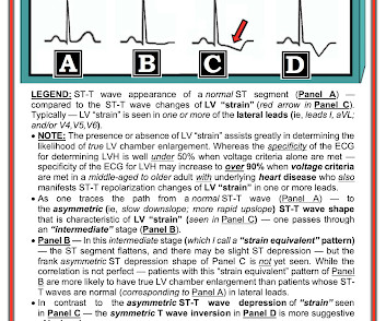
Her vitals signs were remarkable for marked hypertension. would require the ST/S ratio to be 25% for diagnosis of STEMI in LVH. The physician was concerned about STEMI, but also worried that she was overreacting, with the potential that LVH was producing a "STEMI-mimic." Is LVH like left ventricular aneurysm?
The prehospital and ED computer interpretation was inferior STEMI: There’s normal sinus rhythm, first degree AV block and RBBB, normal axis and normal voltages. The paramedic notes called STEMI into question: “EMS disagree with monitor for STEMI callout. Past medical history included diabetes and hypertension.
The Queen of Hearts correctly says: Smith : Why is this ECG which manifests so much ST Elevation NOT a STEMI (even if it were a 60 year old with chest pain)? He was mildly tachycardic (105-110 bpm) and hypertensive (157/92 mm Hg) on arrival. Physician interpretation: "No STEMI." Physician: "No STEMI."
A middle-age woman with h/o hypertension was found down by her husband. Note that they finally have laid to rest the new or presumably new LBBB as a criteria for STEMI. Note that they finally have laid to rest the new or presumably new LBBB as a criteria for STEMI. With ventilations and epinephrine, she regained a pulse.
Written by Pendell Meyers, edits by Steve Smith A man in his 60s with history of hypertension and MI 10 years ago, with PCI, presented to an outside hospital complaining of chest pain that started while mowing the lawn. There is a very small amount of STE in some of the anterior, lateral, and inferior leads which do NOT meet STEMI criteria.
iv ) The findings in Figure-4 could reflect LV aneurysm. C ASE F ollow- U p: I later learned the history in today's case which was that a middle-aged man with diabetes and hypertension who presented to the ED ( E mergency D epartment ) for abdominal pain that had awakened him from sleep. Chest X-Ray was normal.
A 69 year old woman with a history of hypertension presented to the emergency department by EMS for evaluation of chest pain and shortness of breath. There are no Q-waves to suggest old inferior MI, or inferior aneurysm as the etiology of the ST Elevation. Supply-demand mismatch can cause ST Elevation (Type 2 STEMI). Management?
The cath lab was deactivated by cardiologist on arrival at ED because it was "not a STEMI". He carries the diagnoses hyperlipidemia, hypertension, and diabetes. No thoracic aortic hematoma, aneurysm or dissection. Pt received 324 ASA and 2 sprays of nitro with improvement. Cath lab was activated by EMS and transported emergent."
When there are QS-waves, one should always think about LV aneurysm, but ST to QRS ratio and T-wave to QRS ratio are far too large and not compatible with left ventricular aneurysm. 50% of LAD STEMI have Q-waves by one hour. There is some R wave in the lateral precordial leads. Leads V3 and V4 both have 6mm ST elevation.
We organize all of the trending information in your field so you don't have to. Join thousands of users and stay up to date on the latest articles your peers are reading.
You know about us, now we want to get to know you!
Let's personalize your content
Let's get even more personalized
We recognize your account from another site in our network, please click 'Send Email' below to continue with verifying your account and setting a password.


Let's personalize your content