This site uses cookies to improve your experience. To help us insure we adhere to various privacy regulations, please select your country/region of residence. If you do not select a country, we will assume you are from the United States. Select your Cookie Settings or view our Privacy Policy and Terms of Use.
Cookie Settings
Cookies and similar technologies are used on this website for proper function of the website, for tracking performance analytics and for marketing purposes. We and some of our third-party providers may use cookie data for various purposes. Please review the cookie settings below and choose your preference.
Used for the proper function of the website
Used for monitoring website traffic and interactions
Cookie Settings
Cookies and similar technologies are used on this website for proper function of the website, for tracking performance analytics and for marketing purposes. We and some of our third-party providers may use cookie data for various purposes. Please review the cookie settings below and choose your preference.
Strictly Necessary: Used for the proper function of the website
Performance/Analytics: Used for monitoring website traffic and interactions
Aim:This study investigates the prevalence of isolated interventricular membranous septal (IVMS) aneurysms detected via echocardiography and assesses the associated stroke risk without other classical risk factors.Methods:We searched the echocardiography database at Mount Sinai Morningside from January 2017 to September 2023.
IntroductionMycotic aneurysms of paraspinal arteries are a rare finding. Furthermore, knowledge regarding the management of paraspinal mycotic aneurysms and the efficacy of endovascular repair of these lesions is scarce.⁴MethodsWe Stroke: Vascular and Interventional Neurology, Volume 3, Issue S2 , November 1, 2023.
An initial DSA showed a ruptured aneurysm in the second segment of the right posterior inferior cerebellar artery, second segment. However, clinical management of this complication is still debated among experts. [4] He was intubated by EMS on route.
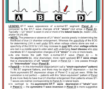
She is somewhat hypertensive, but her vital signs are otherwise normal. However, old MI w/aneurysm morphology (persistent ST-Elevation) can look just like this. While this may be change that is reciprocal to an Acute/Subacute Inferior STEMI, the problem is that LV aneurysm may also manifest with this reciprocal change.
Case Description:A 59-year-old male with history of hypertension, diabetes, Hashimoto’s thyroiditis presented with new, progressive shortness of breath. Coronary angiography revealed a tortuous and extremely aneurysmal RCA, as well as multivessel coronary artery disease (mvCAD) involving LAD, D1, LCx, OM1.
Introduction:Medical treatment of internal carotid artery stenosis consists of treatment of underlying conditions such as hypertension, dyslipidemia, and diabetes mellitus, as well as antiplatelet therapy. Similarly, cerebral aneurysms are known to progress due to hemodynamic effects.
This unique case highlights the diagnostic and therapeutic challenges of a patient with multiple vascular risk factors who suffered from strokes secondary to BHS.MethodsA 79‐year‐old man with a past medical history of peripheral artery disease, abdominal aortic aneurysm, myocardial infarction with drug eluding stents (on dual antiplatelet therapy (DAPT)), (..)
Past medical history included diabetes and hypertension. Peak troponin was a massive 500,000 ng/L, echo showed EF reduced to 20%, and follow up ECG showed LV aneurysm morphology with anterior Q wave and persisting ST elevation. Vitals were normal. There’s normal sinus rhythm, RBBB, normal axis and normal voltages.
New guidelines also: Classify “Elevated BP” between non-elevated BP and hypertension. Measuring eGFR and albuminuria is recommended for assessing kidney disease in all hypertensive patients. Advise increased potassium intake for hypertensive patients.
iv ) The findings in Figure-4 could reflect LV aneurysm. C ASE F ollow- U p: I later learned the history in today's case which was that a middle-aged man with diabetes and hypertension who presented to the ED ( E mergency D epartment ) for abdominal pain that had awakened him from sleep. Chest X-Ray was normal.
When there are QS-waves, one should always think about LV aneurysm, but ST to QRS ratio and T-wave to QRS ratio are far too large and not compatible with left ventricular aneurysm. There is some R wave in the lateral precordial leads. Leads V3 and V4 both have 6mm ST elevation. This ECG shows a lot of "acuity".
He carries the diagnoses hyperlipidemia, hypertension, and diabetes. No thoracic aortic hematoma, aneurysm or dissection. Here is the cardiology note, paraphrased to make it not identifiable: 50-something seen in cardiology consultation today at the request of Dr. XXXXXX for an NSTEMI. CT Angio Chest IMPRESSION 1.
We organize all of the trending information in your field so you don't have to. Join thousands of users and stay up to date on the latest articles your peers are reading.
You know about us, now we want to get to know you!
Let's personalize your content
Let's get even more personalized
We recognize your account from another site in our network, please click 'Send Email' below to continue with verifying your account and setting a password.


Let's personalize your content