This site uses cookies to improve your experience. To help us insure we adhere to various privacy regulations, please select your country/region of residence. If you do not select a country, we will assume you are from the United States. Select your Cookie Settings or view our Privacy Policy and Terms of Use.
Cookie Settings
Cookies and similar technologies are used on this website for proper function of the website, for tracking performance analytics and for marketing purposes. We and some of our third-party providers may use cookie data for various purposes. Please review the cookie settings below and choose your preference.
Used for the proper function of the website
Used for monitoring website traffic and interactions
Cookie Settings
Cookies and similar technologies are used on this website for proper function of the website, for tracking performance analytics and for marketing purposes. We and some of our third-party providers may use cookie data for various purposes. Please review the cookie settings below and choose your preference.
Strictly Necessary: Used for the proper function of the website
Performance/Analytics: Used for monitoring website traffic and interactions
This condition, called atherosclerosis, narrows the arteries, restricting blood flow and increasing the risk of heart attacks and strokes. Increased Risk of Aneurysms : Chronic high blood pressure can weaken the walls of your arteries, leading to bulging areas known as aneurysms.
IntroductionMycotic aneurysms of paraspinal arteries are a rare finding. Furthermore, knowledge regarding the management of paraspinal mycotic aneurysms and the efficacy of endovascular repair of these lesions is scarce.⁴MethodsWe Stroke: Vascular and Interventional Neurology, Volume 3, Issue S2 , November 1, 2023.
Old ‘NSTEMI’ A history of coronaryarterydisease and a stent to the same territory further increases pre-test likelihood of acute coronary occlusion, including in-stent thrombosis. Smith : Old inferior MI with persistent ST Elevation ("inferior aneurysm") has well-formed Q-waves.
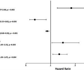
2.40];P<0.001), and aortic aneurysm (odds ratio, 1.64 [95% CI, 1.41–1.92];P<0.001). Genetically predicted BMR may not be causally associated with coronaryarterydisease and ischemic stroke risk. 1.67];P<0.001), atrial fibrillation and flutter (odds ratio, 2.12 [95% CI, 1.87–2.40];P<0.001),
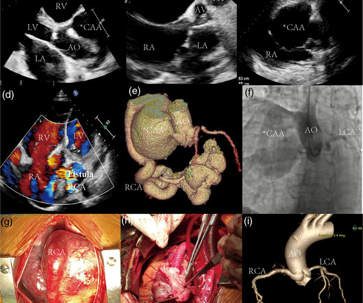
Introduction:Sinus of Valsalva aneurysm (SVA) accounts for 3.5% Her heart failure was due to the fistula as she had no coronaryarterydisease on coronary angiogram. Circulation, Volume 150, Issue Suppl_1 , Page A4118882-A4118882, November 12, 2024. of all congenital cardiac anomalies.
Coronary angiography revealed a tortuous and extremely aneurysmal RCA, as well as multivessel coronaryarterydisease (mvCAD) involving LAD, D1, LCx, OM1. Notably, the LAD had multiple aneurysmal segments and areas of eccentric stenosis upto 90%.Multislice
The patient's heart had significant recovery: Echo : Estimated LVEF 32%, apical wall motion abnormality with diastolic distortion (LV aneurysm), suggestive of old MI. MINOCA: Myocardial Infarction in the Absence of Obstructive CoronaryArteryDisease). 2) overlooked obstructive coronarydisease (e.g.,
Introduction The presence of non-coronary atherosclerosis (NCA) in patients with coronaryarterydisease is associated with a poor prognosis. We have studied whether NCA is also a predictor of poorer outcomes in patients undergoing coronaryartery bypass grafting (CABG).
Atherosclerotic cardiovascular disease (ASCVD), caused by plaque buildup in arterial walls, is one of the leading causes of disability and death worldwide.1,2 1,2 ASCVD causes or contributes to conditions that include coronaryarterydisease (CAD), cerebrovascular disease, and peripheral vascular disease (inclusive of aortic aneurysm).3
In most cases, rather, the culprit is gross ischemia due to myocardial infarction, cardiomyopathy, or advanced coronaryarterydisease. It was postulated that such an ECG feature is associated with advanced myocardial dysfunction, to include left ventricular aneurysm, as the cause of arrhythmia. [7]
There are no Q-waves to suggest old inferior MI, or inferior aneurysm as the etiology of the ST Elevation. The scan showed a bicuspid aortic valve with severe stenosis and coronaryarterydisease. However, there is also significant tachycardia , with heart rate of 116, and known hypoxia.
We organize all of the trending information in your field so you don't have to. Join thousands of users and stay up to date on the latest articles your peers are reading.
You know about us, now we want to get to know you!
Let's personalize your content
Let's get even more personalized
We recognize your account from another site in our network, please click 'Send Email' below to continue with verifying your account and setting a password.


Let's personalize your content