This site uses cookies to improve your experience. To help us insure we adhere to various privacy regulations, please select your country/region of residence. If you do not select a country, we will assume you are from the United States. Select your Cookie Settings or view our Privacy Policy and Terms of Use.
Cookie Settings
Cookies and similar technologies are used on this website for proper function of the website, for tracking performance analytics and for marketing purposes. We and some of our third-party providers may use cookie data for various purposes. Please review the cookie settings below and choose your preference.
Used for the proper function of the website
Used for monitoring website traffic and interactions
Cookie Settings
Cookies and similar technologies are used on this website for proper function of the website, for tracking performance analytics and for marketing purposes. We and some of our third-party providers may use cookie data for various purposes. Please review the cookie settings below and choose your preference.
Strictly Necessary: Used for the proper function of the website
Performance/Analytics: Used for monitoring website traffic and interactions
Coronary aneurysm, a dilated segment of the coronary artery, is a rare condition with a prevalence ranging from 0.02% to 0.2%. According to the current literature, reports of large aneurysms in the left main artery are extremely rare. Due to the high surgical risk, conservative treatment was chosen.
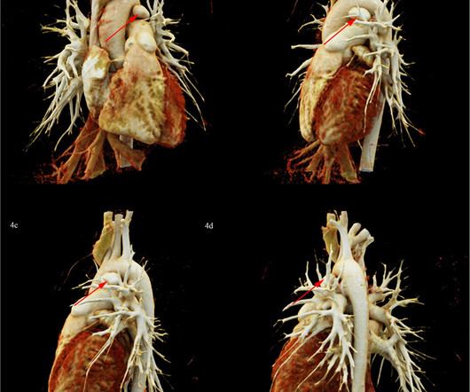
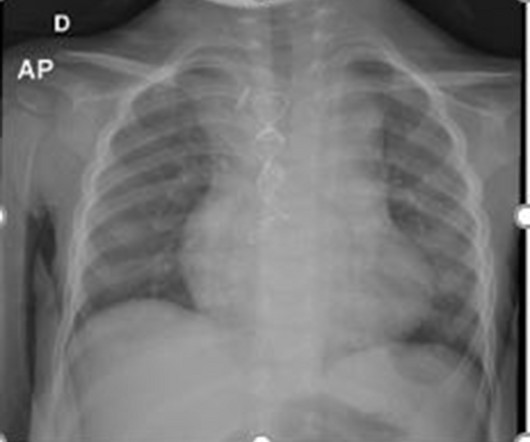
Patent ductus arteriosus (PDA) is one of the most common forms of congenital heart disease (CHD). PDA combined with pulmonary artery aneurysm (PAA) due to IE is rare in children. Infectious endocarditis (IE) is a rare but serious complication of PDA. In this report, we report a rare pediatric PDA case, complicated with PAA due to IE.
Background Bicuspid aortic valve (BAV) is the most common congenital heart defect in adults, often leading to complications such as thoracic aortic aneurysms and aortic stenosis. While BAV is frequently associated with 22q11.2
We present a case of a 22-month-old boy with a hypokinetic and thin-walled aneurysm of the left ventricle apex. Cardiac magnetic resonance imaging revealed an akinetic aneurysm of the LV apex with a full-wall ischemic scar. Determining the origin of the aneurysm is challenging.
Sinus of Valsalva aneurysm (SVA) is an extremely rare condition, and its rupture causes acute symptoms such as chest pain and dyspnea. Ruptured SVA is frequently associated with other congenital defects.
Aortic dissection in pediatrics is an extremely rare condition, which is generally related to predisposing factors such as connective tissue disorders, congenital heart disease and systemic arterial hypertensi.
ABSTRACT Introduction Left atrial appendage aneurysm (LAAA) is a rare congenital cardiac anomaly that involves the progressive dilatation of the left atrial appendage (LAA), predisposing the patient to serious complications such as atrial tachyarrhythmias, life-threatening systemic thromboembolism, and cardiac dysfunction.
Sinus of Valsalva aneurysm (SOVA) is a rare congenital or acquired cardiac defect most often involving the right aortic sinus which is directly related to the interventricular septum. Patients with SOVA may develop conduction system disturbances as a result of direct compression or following operative repair.
Webinar Latin Heart Rounds Series: Rounds on a Patient with Ascending Aortic Aneurysm and Arch Involvement dkaczmarek Wed, 10/11/2023 - 11:22 December 15, 2023 Image Join us on December 15, when experts will provide up-to-date insight on the management of patients with aortic root and ascending aortic aneurysm involving the aortic arch.
Aortopathy encompasses a spectrum of conditions predisposing to dilation, aneurysm, dissection, or rupture of the aorta and other blood vessels. Pathogeneses include connective tissue disorders, smooth muscle contraction disorders, and congenital heart disease, including bicuspid aortic valve, among others. Circulation, Ahead of Print.
It can provide intraluminal fly-through and clipping-plane views which help endovascular assessment of stents, aneurysms, vessel wall irregularities and calcification. The dataset could even be used to generate 3D-printed models of congenital heart disease [2]. Congenit Heart Dis. Congenit Heart Dis. doi: 10.1111/chd.12838.
About 1 in 100 people are born with a bicuspid aortic valve, making it the most common cause of congenital heart disease. A bicuspid aortic valve is a congenital heart defect where the value has two flaps, or cusps, instead of three, so the valve does not open and close properly with each heartbeat.
Introduction:Sinus of Valsalva aneurysm (SVA) accounts for 3.5% of all congenital cardiac anomalies. Aneurysm rupture typically forms a fistula into the right ventricle (60%), right atrium (29%), left atrium (6%), or left ventricle (4%), and rarely into the pericardial cavity (1%).
Coronary angiography revealed a tortuous and extremely aneurysmal RCA, as well as multivessel coronary artery disease (mvCAD) involving LAD, D1, LCx, OM1. Notably, the LAD had multiple aneurysmal segments and areas of eccentric stenosis upto 90%.Multislice CCF can be congenital or acquired and has many variations.
ET – Congenital Cardiac Q&A 6:00 p.m. The program also focuses on reimbursement issues affecting the specialty as a whole. Live Q&A and Discussion Schedule Friday, February 10 4:00 p.m. Live Q&A and Discussion Schedule Friday, February 10 4:00 p.m. ET – 4:15 p.m. ET – Welcome/Introductions 4:15 p.m. ET – 5:15 p.m.
The clinical manifestations of this genetic condition include congenital mydriasis, patent ductus arteriosus, stroke, and/or aortic aneurysms or dissections among many others.
genes implicated in congenital Long QT syndrome, such as SCN5A and KCNQ1). [1-3, It was postulated that such an ECG feature is associated with advanced myocardial dysfunction, to include left ventricular aneurysm, as the cause of arrhythmia. [7]
Spontaneous atrial dissection and atrial aneurysm are extremely rare conditions. This may be the first reported case diagnosed in the fetal period and successfully treated with surgery under extracorporeal circulation in infancy.
An echocardiogram revealed the heart condition that would define much of his early life: severe congenital heart defects. After countless tests, doctors confirmed that it wasnt a seizure, but an aneurysm in his LVOT (left ventricular outflow tract). That test revealed an irregular heart rate, prompting a more in-depth evaluation.
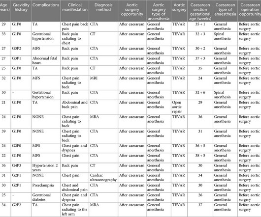
It leads to thickening and loss of elasticity of the arterial wall, and eventually vascular occlusion, aneurysm or dissection formation. TBAD in TA pregnant women is very rare, and the condition is often complicated.
We organize all of the trending information in your field so you don't have to. Join thousands of users and stay up to date on the latest articles your peers are reading.
You know about us, now we want to get to know you!
Let's personalize your content
Let's get even more personalized
We recognize your account from another site in our network, please click 'Send Email' below to continue with verifying your account and setting a password.


Let's personalize your content