This site uses cookies to improve your experience. To help us insure we adhere to various privacy regulations, please select your country/region of residence. If you do not select a country, we will assume you are from the United States. Select your Cookie Settings or view our Privacy Policy and Terms of Use.
Cookie Settings
Cookies and similar technologies are used on this website for proper function of the website, for tracking performance analytics and for marketing purposes. We and some of our third-party providers may use cookie data for various purposes. Please review the cookie settings below and choose your preference.
Used for the proper function of the website
Used for monitoring website traffic and interactions
Cookie Settings
Cookies and similar technologies are used on this website for proper function of the website, for tracking performance analytics and for marketing purposes. We and some of our third-party providers may use cookie data for various purposes. Please review the cookie settings below and choose your preference.
Strictly Necessary: Used for the proper function of the website
Performance/Analytics: Used for monitoring website traffic and interactions
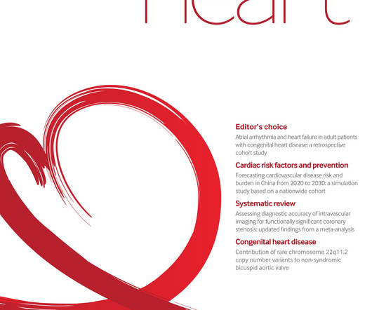
Background Bicuspid aorticvalve (BAV) is the most common congenital heart defect in adults, often leading to complications such as thoracic aorticaneurysms and aorticstenosis. While BAV is frequently associated with 22q11.2
1, 2024 — Researchers at UTHealth Houston have identified genetic variants linked to a rare form of bicuspid aorticvalve disease that affects young adults and can lead to dangerous and potentially life-threatening aortic complications. tim.hodson Wed, 09/04/2024 - 15:53 Sept.
AorticValve Replacement (AVR): AVR is a minimally invasive procedure in which the surgeon makes a small incision and allows the heart to continue to beat during the entire procedure. During a TAVR, the surgeon collapses an artificial aorticvalve and inserts it through a catheter in the chest or leg.
BackgroundSupravalvar aorticstenosis (SVAS) is a characteristic feature of Williams–Beuren syndrome (WBS). Journal of the American Heart Association, Ahead of Print. Its severity varies: ~20% of people with Williams–Beuren syndrome have SVAS requiring surgical intervention, whereas ~35% have no appreciable SVAS.
Mitral valve leaflets seen in open position between the left ventricle and left atrium are thickened. The large aortic regurgitation jet can be seen as a mosaic jet in the left ventricular outflow tract anterior to the anterior mitral leaflet. A portion of the thickened aorticvalve can be seen between the aorta and left ventricle.
There are no Q-waves to suggest old inferior MI, or inferior aneurysm as the etiology of the ST Elevation. Smith : "decompensation" of aorticstenosis might have initiated this entire cascade. What "initiates" the aorticstenosis cascade? No more EKGs were recorded during the patients admission.
We organize all of the trending information in your field so you don't have to. Join thousands of users and stay up to date on the latest articles your peers are reading.
You know about us, now we want to get to know you!
Let's personalize your content
Let's get even more personalized
We recognize your account from another site in our network, please click 'Send Email' below to continue with verifying your account and setting a password.


Let's personalize your content