This site uses cookies to improve your experience. To help us insure we adhere to various privacy regulations, please select your country/region of residence. If you do not select a country, we will assume you are from the United States. Select your Cookie Settings or view our Privacy Policy and Terms of Use.
Cookie Settings
Cookies and similar technologies are used on this website for proper function of the website, for tracking performance analytics and for marketing purposes. We and some of our third-party providers may use cookie data for various purposes. Please review the cookie settings below and choose your preference.
Used for the proper function of the website
Used for monitoring website traffic and interactions
Cookie Settings
Cookies and similar technologies are used on this website for proper function of the website, for tracking performance analytics and for marketing purposes. We and some of our third-party providers may use cookie data for various purposes. Please review the cookie settings below and choose your preference.
Strictly Necessary: Used for the proper function of the website
Performance/Analytics: Used for monitoring website traffic and interactions
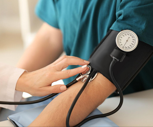
Increased Risk of Aneurysms : Chronic high blood pressure can weaken the walls of your arteries, leading to bulging areas known as aneurysms. If an aneurysm ruptures, it can cause life-threatening internal bleeding. This condition reduces blood flow to the heart, increasing the risk of angina (chest pain) and heart attacks.
There were many comments that it was too late for thrombolytics or that this signified an LV aneurysm, not acute MI. See my formula for differentiating anterior LV aneurysm (that is to say, persistent ST elevation after old MI) from acute anterior STEMI. See my answer below. This is my response: "This is definitely acute or subacute.
A middle-aged man complained of 15 minutes of classic angina that resolved upon arrival to the ED. Although diagnostic of MI, it is highly suspicious for " Old inferior MI with persistent ST Elevation" or "inferior aneurysm morphology" because of the well-formed Q-waves and the flat T-waves. Here is his initial ECG: What do you think?
The patient was referred for an exercise nuclear study and did 11 min on the Bruce protocol without angina or ischaemic ECG changes. Aortic dissection Sinus of Valsalva aneurysm Anomalous coronary artery Unroofed. Physical examination, an ECG, basic laboratories and a chest X-ray were unremarkable.
Coronary angiography revealed a tortuous and extremely aneurysmal RCA, as well as multivessel coronary artery disease (mvCAD) involving LAD, D1, LCx, OM1. Notably, the LAD had multiple aneurysmal segments and areas of eccentric stenosis upto 90%.Multislice CCF can be congenital or acquired and has many variations.
Some case reports have identified IVIg as potential therapy for vasospasm, while others have implicated it as a causative agent in primarily coronary artery vasospasm and the development of atypical angina.
The old ECG has a Q-wave with persistent ST elevation in lead III, and some reciprocal ST depression (typical for aneurysm morphology). This is "Persistent ST elevation after previous MI" or "LV aneurysm morphology". LV aneurysm is very different for inferior vs. anterior MI.
The full thickness infarction with LV aneurysm morphology places him at a higher risk for short and long term complications (e.g., Free wall rupture, VSD, Dresslers Syndrome, chronic CHF, anatomic LV aneurysm, LV thrombus, stroke, etc). No further echocardiograms were available after cath. Teaching points: 1.
Angina is another common symptom due the hypertrophy which causes a coronary supply demand mismatch Symptoms of HCM include syncope/near syncope, which could be precipitated by exertion, hypovolemia, rapid standing, Valsalva manoeuvre, diuretics, vasodilators or arrhythmia. Doppler echo showing LVOT gradient in HCM.
1,2 ASCVD causes or contributes to conditions that include coronary artery disease (CAD), cerebrovascular disease, and peripheral vascular disease (inclusive of aortic aneurysm).3 The benefit was most significant in reducing the incidence of stroke and angina requiring revascularization.35 4 In the U.S. 12 Importantly, colchicine, 0.5
A middle-aged woman had intermittent angina for 48 hours, then onset of constant, crushing chest pain for 1.5 More likely, the patient had crescendo angina, with REVERSIBLE ischemia for 48 hours that only became potentially irreversible (STEMI) at that point in time. Perhaps she will not develop an LV aneurysm.
When there are QS-waves, one should always think about LV aneurysm, but ST to QRS ratio and T-wave to QRS ratio are far too large and not compatible with left ventricular aneurysm. There is some R wave in the lateral precordial leads. Leads V3 and V4 both have 6mm ST elevation. This ECG shows a lot of "acuity".
We organize all of the trending information in your field so you don't have to. Join thousands of users and stay up to date on the latest articles your peers are reading.
You know about us, now we want to get to know you!
Let's personalize your content
Let's get even more personalized
We recognize your account from another site in our network, please click 'Send Email' below to continue with verifying your account and setting a password.


Let's personalize your content