This site uses cookies to improve your experience. To help us insure we adhere to various privacy regulations, please select your country/region of residence. If you do not select a country, we will assume you are from the United States. Select your Cookie Settings or view our Privacy Policy and Terms of Use.
Cookie Settings
Cookies and similar technologies are used on this website for proper function of the website, for tracking performance analytics and for marketing purposes. We and some of our third-party providers may use cookie data for various purposes. Please review the cookie settings below and choose your preference.
Used for the proper function of the website
Used for monitoring website traffic and interactions
Cookie Settings
Cookies and similar technologies are used on this website for proper function of the website, for tracking performance analytics and for marketing purposes. We and some of our third-party providers may use cookie data for various purposes. Please review the cookie settings below and choose your preference.
Strictly Necessary: Used for the proper function of the website
Performance/Analytics: Used for monitoring website traffic and interactions
However, the image quality varies with operator skills as acquiring and interpreting ultrasound images requires extensive training due to the imaging artefacts, the range of acquisition parameters and the variability of patient anatomies.
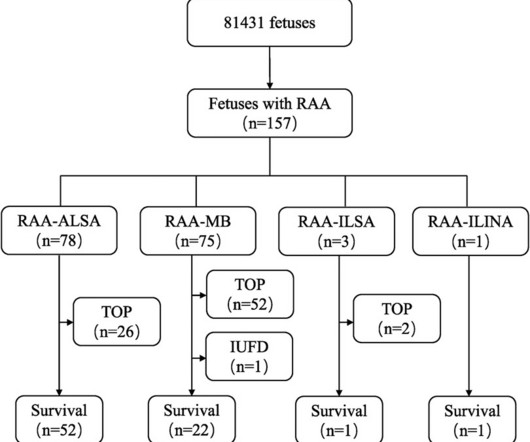
RAA features were characterised by comparing prenatal ultrasound data with anatomical casting results after pregnancy termination or postnatal imaging and surgical intervention to analyse the prognosis and misdiagnoses of fetal RAA.ResultsOf the 157 fetal RAA cases, 50 (31.8%) cases were isolated RAA and 107 (68.2%) cases were nonisolated RAA.
Deep venous anatomy and obstructions can present a multitude of complexities and mechanical challenges. Engineered for the unique demands of venous anatomy and obstructions, the Duo Venous Stent System is comprised of two stents – Duo Hybrid and Duo Extend – of various sizes. It is the third most common cardiovascular disease [2].
a digital health company transforming care through the power of portable, semiconductor-based ultrasound technology and intuitive software, announced today the commercial launch of its third-generation handheld point-of-care ultrasound (POCUS) system, Butterfly iQ3 , which received FDA clearance last month.
Compared to a standard adult population, there are special considerations when planning device implantation in ACHD patients, especially those with complex anatomy.
Intracardiac echocardiography is increasingly utilized for atrial fibrillation (AF) ablation to perform transseptal puncture, monitor for complications and to define left atrial anatomy.
The primary reason is, the LV epicardial lead pacing site was pre-selected by the coronary sinus anatomy. Green: Micra leadless pacemaker; blue: WiSE-CRT system LV endocardial electrode; and red: WiSE-CRT system subcutaneous battery and ultrasound generator. Leadless Ultrasound-Based Cardiac Resynchronization System in Heart Failure.
Bedside ultrasound with no apparent wall motion abnormalities, no pericardial effusion, no right heart strain. First hs troponin I returned 108 minutes after ED arrival and was normal : (12 ng/L) _ No "upstream" P2Y12 were given in the ED ("upstream" means "before the angiogram "defines" the coronary anatomy).
Meanwhile, over the years, ultrasound moved up from the pelvis, abdomen, right into coronary arteries and heart. Intravascular ultrasound-based interventions are being done in coronary artery, in a few cases to avoid contrast in patients with CKD. (We has limited use in deep vision of coronary wall anatomy and histology.
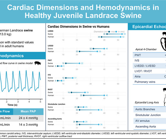
Swine are frequently used as animal model for cardiovascular research, especially in terms of representativity of human anatomy and physiology. Reference values for the most common species used in research are.
Focusing on mitral and tricuspid valve diseases , Capstans treatment combines transcatheter implantation of a folded valve replacement with its X-ray and ultrasound-guided robot to align the low-profile implant with the beating heart valve.
These encompass perceived technical complexities, initial learning curve for practitioners transitioning from femoral to radial access, and considerations regarding access vessel size and patient anatomy variability. Overcoming Technical Hurdles: Mastery of radial access involves employing specific techniques and tools.
These encompass perceived technical complexities, initial learning curve for practitioners transitioning from femoral to radial access, and considerations regarding access vessel size and patient anatomy variability. Overcoming Technical Hurdles: Mastery of radial access involves employing specific techniques and tools.
An intravascular ultrasound was also performed, which was negative for vessel dissection. This case report highlights the modifications in standard techniques, emphasizing the need for specialized skills and strategies to achieve successful outcomes in such cases.
The procedure was initiated via ultrasound‐guided right common femoral access. For patients with complex and unfavorable vascular anatomy for mechanical endo‐vascularization, direct percutaneous carotid puncture can be advantageous, effectively reducing the aforementioned difficulties. ASPECT score 5‐6.
Using machine learning algorithms, InView’s Categorical Hanging Image Protocol(CHIP) automatically organizes images by anatomy and pathology categories to intelligently display or “hang” current and prior studies in a way that significantly improves the efficiency of single or multi-modality image review.
This makes it for instance almost impossible to link the ECG data to the results of Echocardiography (Ultrasound) or CT / MRI imaging. And by relating the ECG data to the heart anatomy we do add a new list of features to the algorithms which are effective in increasing the much needed accuracy in ECG interpretation.
The best course is to wait until the anatomy is defined by angio, then if proceeding to PCI, add Cangrelor (an IV P2Y12 inhibitor) I sent the ECG and clinical information of a 90-year old with chest pain to Dr. McLaren. Widespread ST-depression with reciprocal aVR ST-elevation can be cause by: Heart rate related: tachyarrhythmia (e.g.,
During echocardiography, a transducer transmits the ultrasound beam towards the heart. A good knowledge of the anatomy of the heart is needed for interpretation of images from each view. It is used in the emergency department, at bedside, in the intensive care unit as well as in the operating room.
PMID: 34775811; PMCID: PMC9075358 A bedside ultrasound was performed, shown here: Parasternal short axis view demonstrating inferior LV wall motion akinesis Apical 2 chamber view again demonstrating inferior LV wall akinesis The cath lab was not activated based on the ECG and bedside echo. J Am Heart Assoc. 2021 Dec 7;10(23):e022866.
Beware a negative Bedside ultrasound. Chest Pain in a Male in his 20's; Inferior ST elevation: Inferior lead "early repol" diagnosed. 24 yo woman with chest pain: Is this STEMI? Pericarditis?
Results CARTOSOUND reconstruction was completed using the novel multiplane imaging software platform, allowing for creation of anatomy with minimal movement of the ICE catheter. Discussion Intracardiac ultrasound has become an essential tool in the electrophysiology lab, especially for visualization of intracardiac structures in real time.
First in slow motion with a freeze frame with annotated vessel anatomy, then at normal speed. Nevertheless, the operator performed intravascular ultrasound and saw erupted calcium nodule consistent with plaque erosion. As always I use the same color conventions for vessels as the rest of my angiography guide.
Case continued A bedside cardiac ultrasound revealed grossly preserved left ventricular function, no appreciable wall motion abnormality, pericardial effusion, or obvious valvular abnormality. The terminal part of the T-wave is inverted in lead III, and reciprocally terminally upright in lead aVL.
We organize all of the trending information in your field so you don't have to. Join thousands of users and stay up to date on the latest articles your peers are reading.
You know about us, now we want to get to know you!
Let's personalize your content
Let's get even more personalized
We recognize your account from another site in our network, please click 'Send Email' below to continue with verifying your account and setting a password.


Let's personalize your content