This site uses cookies to improve your experience. To help us insure we adhere to various privacy regulations, please select your country/region of residence. If you do not select a country, we will assume you are from the United States. Select your Cookie Settings or view our Privacy Policy and Terms of Use.
Cookie Settings
Cookies and similar technologies are used on this website for proper function of the website, for tracking performance analytics and for marketing purposes. We and some of our third-party providers may use cookie data for various purposes. Please review the cookie settings below and choose your preference.
Used for the proper function of the website
Used for monitoring website traffic and interactions
Cookie Settings
Cookies and similar technologies are used on this website for proper function of the website, for tracking performance analytics and for marketing purposes. We and some of our third-party providers may use cookie data for various purposes. Please review the cookie settings below and choose your preference.
Strictly Necessary: Used for the proper function of the website
Performance/Analytics: Used for monitoring website traffic and interactions
1 Factors related to congenital heart disease, such as underlying anatomy, surgical repair technique and scars, can all be considered as AA substrates. Of these, AA is one of the leading causes for hospital admissions in ACHD care.
Focal AT at the aorta-mitral annulus conjunction (AMC) is uncommon. Hence, the electrophysiological and ablation target characteristics are poorly described.
Atrioventricular nodal reentry tachycardia (AVNRT), identified as the most prevalent form of regular tachycardia in humans,3 exhibits a strong relationship with the anatomy and physiology of the AV node (AVN) and junction. Despite the temporal distance, Koch's triangle and pyramid remain a repository of unresolved mysteries.
ABSTRACT Atrial flutter (AFL), defined as macro-re-entrant atrial tachycardia, is associated with debilitating symptoms, stroke, heart failure, and increased mortality. Atypical AFL is a heterogenous group of re-entrant atrial tachycardias that most commonly occur in patients with prior heart surgery or catheter ablation.
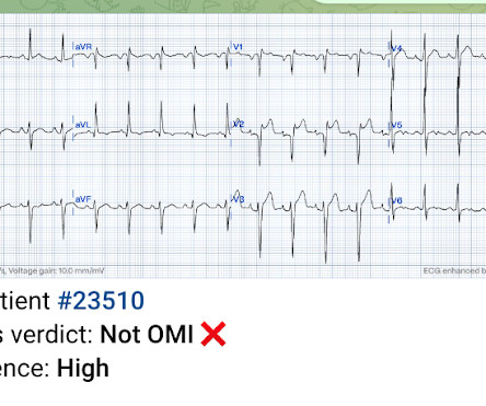
MY Interpretation of Today's Initial ECG: I've labeled key findings in Figure-2 for today's initial ECG: The rhythm is sinus tachycardia at ~105/minute. The sinus tachycardia is a definite concern that something acute may be ongoing. Figure-2: I've labeled t he initial ECG. All intervals ( PR, QRS, QTc ) are normal.
The finding of a fairly regular, wide tachycardia without clear sign of atrial activity ( especially when seen in an acutely symptomatic patient ) — should immediately prompt a diagnosis of VT until proven otherwise. The rhythm looks fairly regular — and atrial activity is absent.
The best course is to wait until the anatomy is defined by angio, then if proceeding to PCI, add Cangrelor (an IV P2Y12 inhibitor) I sent the ECG and clinical information of a 90-year old with chest pain to Dr. McLaren. A slightly prolonged QTc ( although this is difficult to assess given the tachycardia ). A normal PR interval.
To revise the anatomy lessons, this is the external jugular vein and this is the internal jugular vein. One is ventricular tachycardia with regular retrograde activation. Especially, in patients with rheumatic fever, PR interval is prolonged and there is sinus tachycardia. Second is junctional tachycarida.
Anatomy of a Missed LAD Occlusion (classified as a NonSTEMI) See these examples of Septal STEMI: A man in his 50s with "gas pain" A woman in her 70s with chest pain Chest Pain and RBBB. This is sinus tachycardia at a Rate of ~115/minute. Looking for ST depression somewhere, especially in V5, V6 6. wave in V1??
Atrioventricular nodal reentrant tachycardia (AVNRT) is a common supraventricular tachycardia in children and congenital heart disease (CHD) patients. However, in this subgroup ablation remains challenging and experience limited, since anatomy may be atypical and the areas of ablation less predictable or less accessible.
The practice of medicine relies on noninvasive imaging modalities such as computed tomography or magnetic resonance imaging (MRI), which reconstruct noninvasively anatomy of internal organs. Electrocardiographic imaging (ECGI) performs functional imaging. It maps noninvasively the electrical function of the heart.
During observation in the ED the patient had multiple self-terminating runs of Non-Sustained monomorphic Ventricular Tachycardia (NSVT). This patient very likely has some form of idiopathic ventricular tachycardia. Of the ventricular outflow tract tachycardias (RVOT-VT) makes up 80-90%.
It needs a good knowledge of anatomy, physiology of inter & Intra valvular hemodynamics.It It needs a good knowledge of anatomy, physiology of inter & Intra valvular hemodynamics.It Mind you, even an innocuous episode of fever, associated dyspnea, and tachycardia can elevate the mitral gradient and sound a false alarm.
One big chunk of ACS-UA is secondary UA where there is increased demand as in stable angina with tachycardia*. In these patients there is no plaque triggered ACS. For example, in a febrile patient who has associated HT, anemia, etc., You can’t err at the same time , you are not supposed to treat inappropriate as well.
We organize all of the trending information in your field so you don't have to. Join thousands of users and stay up to date on the latest articles your peers are reading.
You know about us, now we want to get to know you!
Let's personalize your content
Let's get even more personalized
We recognize your account from another site in our network, please click 'Send Email' below to continue with verifying your account and setting a password.


Let's personalize your content