This site uses cookies to improve your experience. To help us insure we adhere to various privacy regulations, please select your country/region of residence. If you do not select a country, we will assume you are from the United States. Select your Cookie Settings or view our Privacy Policy and Terms of Use.
Cookie Settings
Cookies and similar technologies are used on this website for proper function of the website, for tracking performance analytics and for marketing purposes. We and some of our third-party providers may use cookie data for various purposes. Please review the cookie settings below and choose your preference.
Used for the proper function of the website
Used for monitoring website traffic and interactions
Cookie Settings
Cookies and similar technologies are used on this website for proper function of the website, for tracking performance analytics and for marketing purposes. We and some of our third-party providers may use cookie data for various purposes. Please review the cookie settings below and choose your preference.
Strictly Necessary: Used for the proper function of the website
Performance/Analytics: Used for monitoring website traffic and interactions
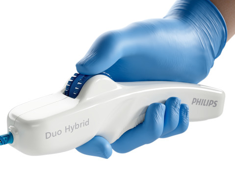
and an investigator in the VIVID study , which contributed to the device’s FDA approval – successfully used the Duo Venous Stent System for the first time outside of a clinical trial. Deep venous anatomy and obstructions can present a multitude of complexities and mechanical challenges. an interventional radiologist with St.
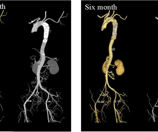
Follow-up CTA scans at one- and six-month post-operation showed that the aortic stent was well-positioned, with no visible primary lesion. After a comprehensive, multidimensional evaluation of the patient's medical history, CTA, and esophagography, we successfully performed TEVAR procedure.
a company focused on reducing the risk of stroke and its devastating impact, today announced that it has launched its Tapered ENROUTE Transcarotid Stent System to hospitals in the United States. New tapered configurations for our ENROUTE Transcarotid Stent System build upon the robust portfolio of Silk Road’s carotid solutions.
announced that the first patients have been enrolled in the Gore VBX FORWARD Clinical Study ( NCT05811364 ), a global prospective, multicenter, randomized controlled trial to compare the GORE VIABAHN VBX Balloon Expandable Endoprosthesis (VBX Stent Graft) to bare metal stenting for patients with complex iliac occlusive disease. "Our
Hes team have recently developed a new Coronary Artery Tree description and Lesion EvaluaTion (CatLet) angiographic scoring system, which is capable of accounting for the variability in coronary anatomy, and.
In-silico evaluation (computer simulated analysis in a virtual stroke model) has the potential to test and optimize large number of stent-retriever design variations in a relatively time and cost-effective manner. Gravity Medical Technology’s SuperNova Stent-retriever was the device under investigation.
In‐silico evaluation (computer simulated analysis in a virtual stroke model) has the potential to test and optimize large number of stent‐retriever design variations in a relatively time and cost‐effective manner. Further, new design concepts can be tested across multiple anatomical scenarios.
Such medium or distal arterial segments have not been assessed with respect to thrombectomy devices used during endovascular therapy. Arterial diameters were measured at all these sites.
Furthermore, it aids in planning and conducting safe aortic intervention and assists in deciding on single- or two-staged stent graft procedures. It underscores the value of preoperative CT councils and provides crucial information for interpreting the results.
First hs troponin I returned 108 minutes after ED arrival and was normal : (12 ng/L) _ No "upstream" P2Y12 were given in the ED ("upstream" means "before the angiogram "defines" the coronary anatomy). Here are other very interesting posts: Wellens' syndrome: to stent or not?
Diagnostic cardiac catheterization may be needed especially in tetralogy of Fallot with pulmonary atresia, to assess the pulmonary anatomy, including size and distribution of peripheral pulmonary arteries. Stenting of the patent ductus arteriosus can be considered in neonatal period for improving oxygen saturation till corrective surgery.
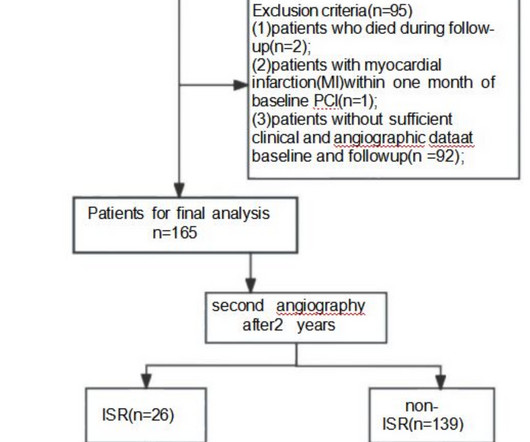
Intra-procedural data included access route, coronary anatomy, lesion complexity, number of stents deployed, door-to-balloon time for primary PCI, and any intra-procedural complications. and the average number of stents 2.6. The radial approach was used in 544/567 (95.94%), the average SYNTAX score was 34.8 ± 9.6,
She was taken to the cath lab, where she was found to have 100% in-stent restenosis of the proximal LAD. Prompt cath to define the anatomy should be expedited. About 45 minutes after the second EKG, the patient was found in cardiac arrest. She was worked as a full code, and ROSC was achieved.
And finally, after placement of a stent in the LAD: Before and after: (Unfortunately, this resulted in the "jailing" of the septal branches behind the stent and probably some degree of plaque shift which is why they do not opacify well in the "after" shot.
has limited use in deep vision of coronary wall anatomy and histology. Now is the era of Optics If a torch light can illuminate and give us vision in absolute darkness ,how about acquiring a deep vision with scattered light ie photons. Jnana-Chakshush ,third eye of Hindu God Shiva ?) As of now it has no role to play in catheter guidance.
After guidewire crossing, balloon angioplasty was performed, and a drug-eluting stent was deployed. The left circumflex had 80% proximal stenosis with minimal luminal irregularities in the mid to distal portion. An intravascular ultrasound was also performed, which was negative for vessel dissection.
For example, interventional cardiologists can benefit from simulation labs that mimic complex cardiovascular procedures, allowing them to practice catheter insertion techniques or stent placements before applying them in a live setting.
Two adjunctive modifications of CE ‐ balloon remodeling techniques (BRT) and stent‐assisted coiling (SAC) ‐ have been utilized to facilitate occlusion of BTAs of variable anatomies/morphologies, sizes, and rupture status. Stents approved by FDA after 2014 (used in 13 cases) had a greater rate of retreatment (46.2% vs. 10.7%).
2017 ) Clinical implication of such coronary anomalies Apart from angiographic surprises, these anomalous coronary arteries may under-perfuse the ventricle and present as unexplained cardiomyopathy , until we realize the anatomical errors in coronary anatomy. Annu Rev Physiol. Some unanswered queries 1.
We aimed to develop a 3D printed ICAD model including realistic features to provide an optimal simulation phantom for research and training purposes.MethodsStereolithography 3D printing technique was used to create a resin neurovascular model based on vascular anatomies extracted from anonymized CTA images.
For example, interventional cardiologists can benefit from simulation labs that mimic complex cardiovascular procedures, allowing them to practice catheter insertion techniques or stent placements before applying them in a live setting.
60-something with h/o MI and stents presented with chest pain radiating to the back and nausea/vomiting. It was stented. The patient had a p rior h istory of MI + stents. Time zero What do you think? There is inferior ST elevation. Is it normal variant? Is it ischemic (OMI)? Pericarditis?
They found 100% acute mid-LAD Occlusion MI, stented with excellent angiographic result. Although I do not see much difference between the ECGs, for some reason (perhaps ongoing pain or rising troponins) the case was reevaluated at this time and the decision was made to perform cath.
They found an acute lesion of the LAD at the site of the prior stents, including 70% proximal LAD lesion and 95% mid-LAD stenosis with TIMI 3 flow at the time of cath. The LAD lesion was acute and required 3 stents to restore flow. because if it does, then urgent cath to define the anatomy is clearly indicated.
Compare to the anatomy after stenting: The lower of the 2 now easily seen branches is the circumflex, now with excellent flow. Here is his angiogram: This shot shows that the left circumflex (LCx) is occluded at the ostium (origin). This is seen just millimeters beyond the tip of the catheter. The patient recovered well.
There is new data showing better outcomes when bystander lesions (non-culprit) are stented. == MY Comment by K EN G RAUER, MD ( 8/28/2020 ): == Dr. Smith highlights a number of important lessons to be learned from today’s case. Initial priorities in this patient were clearly to determine the anatomy — and reestablish coronary perfusion.
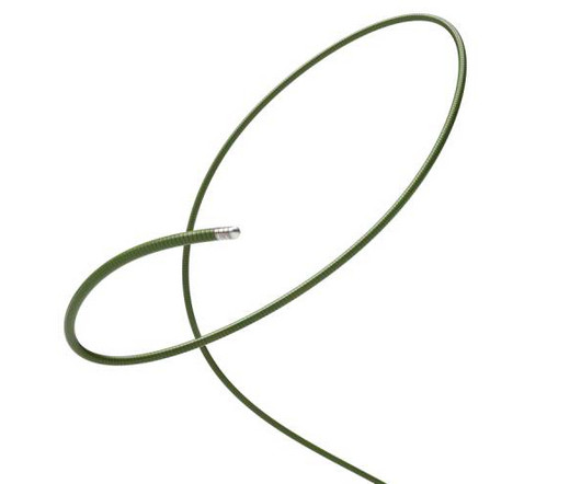
Guidewires are the backbone of the aortic procedure, facilitating tracking through patient anatomy and delivering the stent graft to the landing zone. The ideal wire is a balance of deliverability with patient safety,” said Patrick Muck, MD, vascular surgeon, TriHealth Heart Institute.
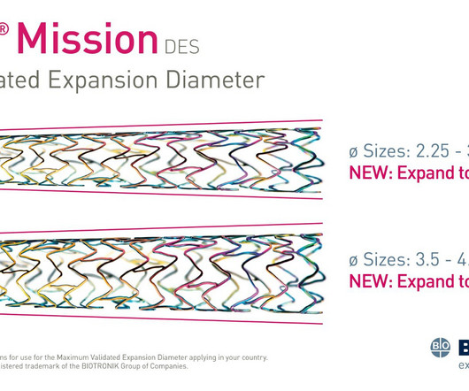
mtaschetta-millane Tue, 07/02/2024 - 09:45 July 2, 2024 — Biotronik announced the availability of an expanded Maximum Allowed Diameters (MAD) range for the Orsiro Mission Drug Eluting Stent (DES). Diameters 2.25 mm to 3.00 mm of the Orsiro Mission DES can now be extended up to 4.0mm, while diameters 3.5 mm and 4.0 mm can reach up to 5.0
He reports this was similar to how he felt when he had his heart attack 4 years prior, now s/p 4 stents. hours: The very suggestive history for an acute event + unrelieved CP alone ( even without ECG abnormalities ) is indication for prompt cath to determine the anatomy. He states that it feels like burning and pressure, like GERD.
The report describes heavy plaque in the proximal RCA by IVUS, but no lesions in the previously occluded RPL branch and no stent was deployed. She underwent coronary angiography which showed thrombotic occlusion of an RPL branch s/p aspiration thrombectomy. It is consistent with an inferior LV aneurysm. No repeat ECGs were obtained.
The RCA was stented successfully with TIMI III flow noted post-procedure and the patient has done well with a post-PCI TTE demonstrating good LVEF and no wall motion abnormality. Angiography was performed and found a normal LAD, a large co-dominant LCX, and 95% disease at the mid-RCA.
We organize all of the trending information in your field so you don't have to. Join thousands of users and stay up to date on the latest articles your peers are reading.
You know about us, now we want to get to know you!
Let's personalize your content
Let's get even more personalized
We recognize your account from another site in our network, please click 'Send Email' below to continue with verifying your account and setting a password.


Let's personalize your content