This site uses cookies to improve your experience. To help us insure we adhere to various privacy regulations, please select your country/region of residence. If you do not select a country, we will assume you are from the United States. Select your Cookie Settings or view our Privacy Policy and Terms of Use.
Cookie Settings
Cookies and similar technologies are used on this website for proper function of the website, for tracking performance analytics and for marketing purposes. We and some of our third-party providers may use cookie data for various purposes. Please review the cookie settings below and choose your preference.
Used for the proper function of the website
Used for monitoring website traffic and interactions
Cookie Settings
Cookies and similar technologies are used on this website for proper function of the website, for tracking performance analytics and for marketing purposes. We and some of our third-party providers may use cookie data for various purposes. Please review the cookie settings below and choose your preference.
Strictly Necessary: Used for the proper function of the website
Performance/Analytics: Used for monitoring website traffic and interactions
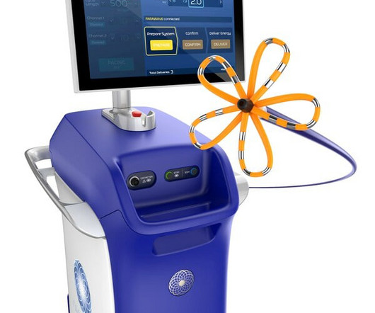
All patients were pre-TAVR assessed by transthoracic echocardiography and computed tomography of the aortic valve (AV) and relevant left cardiac and vascular anatomy. Nanjing) to evaluate its safety and efficacy.Methods130 high risk patients with symptomatic severe AS from 11 institutions were treated with the novel Xcor system.
CTAs were reviewed to assess the carotid stenosis and the anatomy of the aortic arch. Unlimited propensity score matching was performed to balance baseline characteristics between patients with bovine and normal anatomies. Among the 65 patients included, 47 (74.6%) were male, with a median age of 70 [IQR:65.0-76.0]
We aimed to develop a 3D printed ICAD model including realistic features to provide an optimal simulation phantom for research and training purposes.MethodsStereolithography 3D printing technique was used to create a resin neurovascular model based on vascular anatomies extracted from anonymized CTA images.
BACKGROUND:Data concerning the outcomes of transcatheter aortic valve replacement in type 0 bicuspid aortic stenosis (AS) are scarce. Circulation: Cardiovascular Interventions, Ahead of Print. Self-expanding transcatheter heart valves were used in the majority of patients (n=1160; 91.4%). Poverall=0.522; 1 year: 10% versus 2.3%
First hs troponin I returned 108 minutes after ED arrival and was normal : (12 ng/L) _ No "upstream" P2Y12 were given in the ED ("upstream" means "before the angiogram "defines" the coronary anatomy). Pattern B — was the more common form in the original Wellens’ report.
Additional real-world data from more than 17,000 patients in the MANIFEST-17K registry demonstrated continued real-world safety of the system, with no reports of permanent phrenic nerve palsy, pulmonary vein stenosis or esophageal injury. director of electrophysiology, Mount Sinai Fuster Heart Hospital , New York.
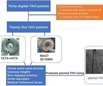
Methods Thirty patients with severe aortic stenosis were prospectively enrolled. The anatomical properties of the aortic root anatomy, including the perimeter and area of the virtual aortic valve annulus and coronary heights, were determined from 3D whole-heart MRI and cardiac CTA (CCTA) images, respectively.
Due to the dramatically improved survival of children with congenital heart disease over the last 5 decades, there has been a steady increase in the prevalence of adults with congenital heart disease, which necessitates that clinicians become familiar with the anatomy and the evaluation of right ventricular outflow tract and PV anomalies.
If you’ve been diagnosed with aortic stenosis, you might have come across the term TAVR. Understanding Aortic Stenosis The aortic valve regulates blood flow from your heart’s main pumping chamber to the rest of your body. In aortic stenosis, the valve leaflets stiffen and narrow, restricting blood flow.
That is, pulmonary artery is transposed over to the right ventricle, and aorta over to the left ventricle, so that normal anatomy is restored. Graft material has the disadvantage that it will not grow as the baby grows and can lead to supravalvar pulmonary stenosis later, one of the delayed complications of arterial switch.
Diagnostic cardiac catheterization may be needed especially in tetralogy of Fallot with pulmonary atresia, to assess the pulmonary anatomy, including size and distribution of peripheral pulmonary arteries. Hemocontration is due to the diuresis following contrast angiography, which can be prevented by adequate pre and post procedure hydration.
Methods We present the first described case of using two leadless pacing systems manufactured by separate companies implanted within the same patient to provide atrial and ventricular pacing due to complex congenital anatomy.
The left circumflex had 80% proximal stenosis with minimal luminal irregularities in the mid to distal portion. After guidewire crossing, balloon angioplasty was performed, and a drug-eluting stent was deployed. An intravascular ultrasound was also performed, which was negative for vessel dissection.
The best course is to wait until the anatomy is defined by angio, then if proceeding to PCI, add Cangrelor (an IV P2Y12 inhibitor) I sent the ECG and clinical information of a 90-year old with chest pain to Dr. McLaren. 2 cases of Aortic Stenosis: Diffuse Subendocardial Ischemia on the ECG. His response: “subendocardial ischemia.
The cath lab was activated: Result: Thrombotic 95% stenosis at the ostium of a small LPL2 with 70% stenosis at the LPL2/LPDA bifurcation in the distal/AV groove Cx Tubular 70% stenosis in the mid-circumflex. (In In other words, inferior MI with some posterior involvement). It was stented.
To revise the anatomy lessons, this is the external jugular vein and this is the internal jugular vein. The Y descent is shallow in tricuspid stenosis, and absent in cardiac tamponade. Right atrial hypertrophy as in tricuspid stenosis, pulmonary stenosis and pulmonary hypertension.
This is essentially pathognomonic for Occlusion MI of the anterior and apical walls (de Winter pattern more likely represents ~99% thrombotic stenosis with just a trickle of flow, but essentially the same overall event, with progression to full occlusion extremely likely).
90% stenosis of the proximal ramus intermedius, pre procedure TIMI II flow The ramus intermedius is a normal variant on coronary anatomy that arises between the LAD and LCX. Serum troponin I level just before the cardiac catheterization procedure was 16.69 Its course is variable, often supplying the lateral wall of the LV.
For example, inferior OMI with concomitant critical stenosis produces a combined pattern ( Aslanger’s pattern ) with inferior STE and subendocardial ischemia · occlusion of two infarct-related arteries simultaneously ("co-culprits") In this case there were two infarct-related arteries.
A good knowledge of the anatomy of the heart is needed for interpretation of images from each view. Planimetry of mitral valve area can be obtained in parasternal short axis view in case of mitral stenosis. Parasternal views are often obtained first, followed by apical, subcostal, and suprasternal.
Here I annotate it: This shows 100% occluded circumflex (red arrow) and a 90% stenosis of the LAD (Yellow arrow). The LAD was thought to be not thrombotic, but a chronic tight stenosis. Initial priorities in this patient were clearly to determine the anatomy — and reestablish coronary perfusion.
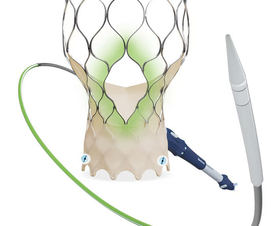
milla1cf Thu, 03/28/2024 - 07:30 March 28, 2024 — Medtronic plc, a global leader in healthcare technology, announced that the United States Food and Drug Administration ( FDA ) has approved the Evolut FX+ transcatheter aortic valve replacement (TAVR) system for the treatment of symptomatic severe aortic stenosis. Aortic stenosis.
The anatomy and lead placement create very small voltage compared to the other main coronary distributions. The American Journal of Cardiology 2010 1,500 consecutive patients with complete occlusion or near occlusion (greater than 90% stenosis with TIMI less than 3) were identified post-hoc from a prospective PCI database. EF was 55%.
All participants underwent coronary computed tomography angiography (CCTA), a non-invasive imaging technique used to assess coronary artery anatomy and stenosis, at baseline and at 12 months. A total of 84 participants were enrolled, of which 72 completed the trial, with participants randomized to receive either colchicine 0.5
The CaRi-Plaque technology supports non-invasive analysis of coronary anatomy and pathology from routine coronary computed tomography angiography (CCTA) scans to determine the presence, extent and severity of coronary plaques and luminal stenosis (narrowing of arteries).
The CoreValve and next-generation Evolut TAVR systems are used in TAVR procedures for patients with symptomatic severe aortic stenosis (AS), which are less invasive and typically result in a quicker recovery time than open heart surgery.i The natural history and rate of progression of aortic stenosis. iii Am Heart Assoc. 120.018816.
All participants underwent coronary computed tomography angiography (CCTA), a non-invasive imaging technique used to assess coronary artery anatomy and stenosis, at baseline and at 12 months. A total of 84 participants were enrolled, of which 72 completed the trial, with participants randomized to receive either colchicine 0.5
TriClip leverages the same clip-based technology as Abbott's leading MitraClip device – which has treated more than 200,000 people with leaky mitral valves (mitral regurgitation) – but was specifically designed to treat the tricuspid valve's complex anatomy. The device has already been used to treat more than 10,000 people with TR.
They found an acute lesion of the LAD at the site of the prior stents, including 70% proximal LAD lesion and 95% mid-LAD stenosis with TIMI 3 flow at the time of cath. because if it does, then urgent cath to define the anatomy is clearly indicated. They took him almost immediately for catheterization.
We organize all of the trending information in your field so you don't have to. Join thousands of users and stay up to date on the latest articles your peers are reading.
You know about us, now we want to get to know you!
Let's personalize your content
Let's get even more personalized
We recognize your account from another site in our network, please click 'Send Email' below to continue with verifying your account and setting a password.


Let's personalize your content