This site uses cookies to improve your experience. To help us insure we adhere to various privacy regulations, please select your country/region of residence. If you do not select a country, we will assume you are from the United States. Select your Cookie Settings or view our Privacy Policy and Terms of Use.
Cookie Settings
Cookies and similar technologies are used on this website for proper function of the website, for tracking performance analytics and for marketing purposes. We and some of our third-party providers may use cookie data for various purposes. Please review the cookie settings below and choose your preference.
Used for the proper function of the website
Used for monitoring website traffic and interactions
Cookie Settings
Cookies and similar technologies are used on this website for proper function of the website, for tracking performance analytics and for marketing purposes. We and some of our third-party providers may use cookie data for various purposes. Please review the cookie settings below and choose your preference.
Strictly Necessary: Used for the proper function of the website
Performance/Analytics: Used for monitoring website traffic and interactions
6, 2025 Medtronic plc hasannounced it received CE ( Conformit Europenne ) Mark for the Harmony Transcatheter Pulmonary Valve (TPV) System, a minimally invasive alternative to open-heart surgery for congenital heart disease patients with native or surgically repaired right ventricular outflow tract (RVOT).
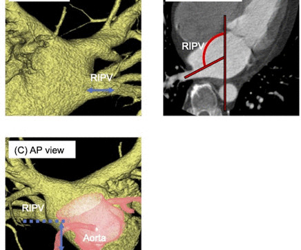
No data have been reported on cooling characteristics and the impact of variant pulmonary vein (PV) anatomy on atrial fibrillation (AF) recurrences after POLARx cryoballoon (CB) ablation.
The Guidezilla guide extension catheter (GEC) with a larger diameter and extended length is widely used in challenging coronary anatomy. However, the constrained accessibility of dedicated catheters has impeded the potential benefits of standard CDT in developing countries.
Studies estimate that severe regurgitation occurs in up to 4% of individuals 75 and older and is often linked with conditions like heart failure, atrial fibrillation or pulmonary hypertension. A TEER procedure, which inserts clips in the valve to reduce leakage, works well for many, but is less effective for those with complex valve anatomy.
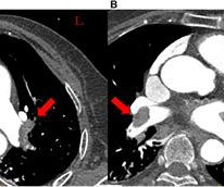
Hemothorax caused by a right intercostal artery (ICA) injury behind the left atrium (LA) is a potentially fatal complication during pulmonary vein isolation. However, their anatomical relationship has not been fully elucidated.
a powerful and advanced mechanical thrombectomy system for the removal of venous thrombus and the treatment of pulmonary embolism (PE), effectively completing Penumbras VTE platform. The Element Vascular Access System is compatible with Lightning Flash 2.0, The flexibility and torqueability of the Lightning Flash2.0
The pivotal trial will study the Vertex Pulmonary Embolectomy System, which incorporates Jupiter’s Endoportal Control platform technology into an endovascular procedure intended to treat Acute Pulmonary Embolism (PE) with an unprecedented level of control and precision. pivotal study (NCT06576427).
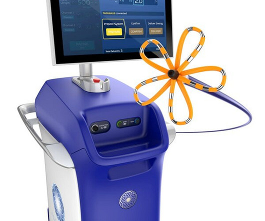
A company statement reported that its PFA System is indicated for the isolation of pulmonary veins in the treatment of drug-refractory, recurrent, symptomatic, paroxysmal (i.e., The FARAPULSE PFA System is indicated for the isolation of pulmonary veins in the treatment of drug-refractory, recurrent, symptomatic, paroxysmal (i.e.,
The trial showed the Volt PFA System achieved pulmonary vein isolation (PVI) the method of destroying tissue causing a patient's AFib in 99.1% As a result, PFA can reduce the risk of damaging adjacent tissue in patients with complex disease or anatomy. 3 Following approval, initial cases were completed by Prof.
Pre-operatively, the controlled dilation may help address unique anatomies. To help navigate the unique anatomy of each patient, exGraft also includes radiopaque markers, which help ensure accurate identification of the graft in the post-implantation period.
Chronic Pulmonary Disease Lung diseases like chronic obstructive pulmonary disease (COPD) can lead to pulmonary hypertension, which in turn can cause the right side of the heart to enlarge, a condition known as cor pulmonale.
“Single-shot” pulsed field ablation (PFA) catheters are challenged by the need for serial re-positioning and rotation, difficulties with easily accommodating varying PV anatomies, and the need for a second catheter to perform electroanatomical mapping.
Chance of precipitating a cyanotic spell are more when pulmonary angiography is attempted through the already narrow right ventricular outflow tract. initial shunt surgery is an option to allow the pulmonary artery branches to grow in size and for a later complete repair of tetralogy of Fallot [1]. If McGoon’s ratio is below 0.8,
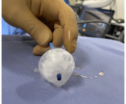
Method A tissue-based pulmonary vein model was constructed from porcine myocardial tissue and placed on a stage designed to simulate pulmonary vein anatomy and venous flow. A temperature sensor was set behind the muscle and cryoballoon ablation was performed after confirming the occlusion of pulmonary vein with cryoballoon.
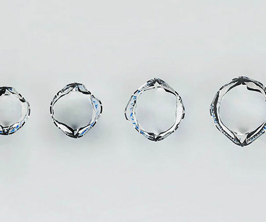
This novel cryoballoon with adjustable size and low compliance successfully achieves pulmonary vein isolation to treat paroxysmal atrial fibrillation (PAF), providing more options for patients with PAF. This cryoballoon system was proven to be safe and effective for treatment of patients with drug refractory or drug intolerant PAF.
Background The limited availability of balloon sizes for cryoballoon leads to anatomical limitations for pulmonary vein (PV) isolation. Search keywords included ‘atrial fibrillation’, ‘cryoballoon ablation’ and ‘anatomy’ Results Overall, 243 articles were identified.
Due to the dramatically improved survival of children with congenital heart disease over the last 5 decades, there has been a steady increase in the prevalence of adults with congenital heart disease, which necessitates that clinicians become familiar with the anatomy and the evaluation of right ventricular outflow tract and PV anomalies.
Unfortunately, the traditional pulmonary valves have a fixed diameter that can’t match the size of the child’s heart over time. The prototype valve has two leaflets crafted from a polymer material that has a well-established history of being used as a leaflet for pediatric pulmonary valves.
Genetically predicted shorter leukocyte telomere length was causally linked to smaller ventricular cavity sizes including left ventricular end‐systolic volume, left ventricular end‐diastolic volume, lower left ventricular mass, and pulmonary artery. However, the direct causality of these relationships has not been definitively established.
That is, right ventricle is connecting to aorta, and left ventricle to pulmonary artery. That is, pulmonary artery is transposed over to the right ventricle, and aorta over to the left ventricle, so that normal anatomy is restored. In simple terms, arterial switch is restoring the normal connections.
According to the company, the Duo system adds high resolution thoracic clinical capability, suitable for screening lung cancer, COVID-19 and other pulmonary diseases. In addition, Arineta Cardiac Imaging announced FDA 510(k) clearance of its SpotLight and SpotLight Duo family of cardiovascular CT scanners at RSNA23. Clinical applications.
Methods We present the first described case of using two leadless pacing systems manufactured by separate companies implanted within the same patient to provide atrial and ventricular pacing due to complex congenital anatomy.
This possiblity depends on the pre-procedural anatomy, the placement of the clips, and the resulting changes in mitral valve dynamics. However, case studies and operator experiences suggest that jet redirection can occur, particularly with suboptimal clip positioning or in complex anatomies. Implication of new onset eccentric jet 1.Eccentric
Untreated, it may result in heart failure due to volume loading of the left heart, pulmonary hypertension, and infective endarteritis. Background:The persistently patent arterial duct accounts for ~12% of congenital heart lesions. Type A ducts were more frequent in the AA group (90% vs. 72%).
The pulmonary semilunar valve is between the right ventricle and the pulmonary trunk. Several major arteries and veins include: The pulmonary artery transports blood with low levels of oxygen and high levels of carbon dioxide to the lungs. Like the heart chambers, there are four heart valves between each of the chambers.
To revise the anatomy lessons, this is the external jugular vein and this is the internal jugular vein. Right atrial hypertrophy as in tricuspid stenosis, pulmonary stenosis and pulmonary hypertension. But in a VSD with pulmonary hypertension A wave is not prominent.
A good knowledge of the anatomy of the heart is needed for interpretation of images from each view. The aorta, right ventricular outflow tract and pulmonary artery up to its bifurcation is imaged in the upward angulation shown in the left panel. Colour flow shows the flow in pulmonary artery.
From there, the right ventricle pumps the blood to the lungs via the pulmonary arteries for reoxygenation. Blood Re-enters Systemic Circulation Once deoxygenated blood reaches the right atrium, it flows into the right ventricle.
CT angiogram chest: no aortic dissection or pulmonary embolism. Serial chest xrays: progressive bilateral pulmonary edema. He spent several days in the PICU, undergoing workup including: Serial troponins: rising from 5,700 ng/L (unknown if I or T) to greater than 25,000 ng/L (greater than the lab's upper limit of reporting).
Studies estimate that severe regurgitation occurs in up to 4% of individuals 75 and older and is often linked with conditions like heart failure, atrial fibrillation or pulmonary hypertension. A TEER procedure, which inserts clips in the valve to reduce leakage, works well for many, but is less effective for those with complex valve anatomy.
His initial high sensitivity troponin I returned at 1300 ng/L and given that his cardiac workup was otherwise unremarkable, a CT was obtained to evaluate for pulmonary embolism and aortic aneurysm or dissection but this too was unrevealing. Also: electrical instability, pulmonary edema, or hypotension. Another EKG was also obtained.
We organize all of the trending information in your field so you don't have to. Join thousands of users and stay up to date on the latest articles your peers are reading.
You know about us, now we want to get to know you!
Let's personalize your content
Let's get even more personalized
We recognize your account from another site in our network, please click 'Send Email' below to continue with verifying your account and setting a password.


Let's personalize your content