This site uses cookies to improve your experience. To help us insure we adhere to various privacy regulations, please select your country/region of residence. If you do not select a country, we will assume you are from the United States. Select your Cookie Settings or view our Privacy Policy and Terms of Use.
Cookie Settings
Cookies and similar technologies are used on this website for proper function of the website, for tracking performance analytics and for marketing purposes. We and some of our third-party providers may use cookie data for various purposes. Please review the cookie settings below and choose your preference.
Used for the proper function of the website
Used for monitoring website traffic and interactions
Cookie Settings
Cookies and similar technologies are used on this website for proper function of the website, for tracking performance analytics and for marketing purposes. We and some of our third-party providers may use cookie data for various purposes. Please review the cookie settings below and choose your preference.
Strictly Necessary: Used for the proper function of the website
Performance/Analytics: Used for monitoring website traffic and interactions
The registry will collect multi-site, real-world information on how the Plaque Analysis product provides enhanced patient insights, empowering physicians and helping to inform their medical management decisions for patients with suspected coronary artery disease (CAD). 2 “Data from the DECODE study shows the value of using Plaque Analysis.
mg)has potential to directly reduce inflammation, which plays a substantial role in the formation and progression of atherosclerotic plaque leading to heart disease, said Matthew J. mg improved several measures of plaque volume changes over a period of 12 months in patients with stable coronary artery disease, Dr. Budoff continued.
The CaRi-Plaque technology supports non-invasive analysis of coronary anatomy and pathology from routine coronary computed tomography angiography (CCTA) scans to determine the presence, extent and severity of coronary plaques and luminal stenosis (narrowing of arteries). But with AI, we can change that.
a leader in non-invasive artificial intelligence (AI) heart care solutions, announced that the data from its REVEALPLAQUE study , highlighting the accuracy of its Plaque Analysis, was published in the European Heart Journal Cardiovascular Imaging. milla1cf Thu, 05/23/2024 - 08:00 May 23, 2024 — HeartFlow, Inc. ,
The bovine anatomical variation may alter the blood flow dynamics, potentially contributing to the formation and progression of carotid plaques. CTAs were reviewed to assess the carotid stenosis and the anatomy of the aortic arch. Among the 65 patients included, 47 (74.6%) were male, with a median age of 70 [IQR:65.0-76.0]
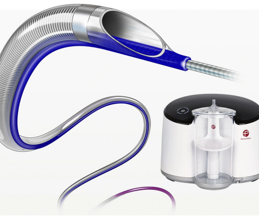
Now commercially available in Europe, CAT RX is designed to navigate tortuous coronary anatomy while maintaining sustained mechanical aspiration with the Penumbra ENGINE. Traditional modalities utilizing syringe aspiration suffer from diminished vacuum once fluid enters the system.
mg)has potential to directly reduce inflammation, which plays a substantial role in the formation and progression of atherosclerotic plaque leading to heart disease, said Matthew J. mg improved several measures of plaque volume changes over a period of 12 months in patients with stable coronary artery disease, Dr. Budoff continued.
First hs troponin I returned 108 minutes after ED arrival and was normal : (12 ng/L) _ No "upstream" P2Y12 were given in the ED ("upstream" means "before the angiogram "defines" the coronary anatomy). Angiography : --Culprit for the patient's unstable angina/Wellen syndrome is a ruptured plaque in the mid LAD. --As
This launch expands upon the company’s prior ENROUTE Transcarotid Stent System, offering additional configurations to better tailor the Transcarotid Artery Revascularization (TCAR) procedure to patient anatomy. The stent system was purpose-built for TCAR with a short delivery system for ergonomic and precise stent delivery.
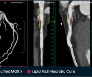
tim.hodson Tue, 10/01/2024 - 10:50 PHOTO CAPTION: The Elucid PlaqueIQ user interface is a fully interactive visualization of the patient’s coronary anatomy, showing specific plaque type and amount across various views to inform physician assessment of risk and patient-specific treatment pathway.
It is generally believed it is more of a mechanical plaque lesion. RCA and LCX Wellens do occur, making this entity’s perceived unique importance less certain 3. How common is thrombosis in the culprit artery of Wellen syndrome ? However by no means, we can say thrombosis do not occur. Is there a benign face of Wellen syndrome ?
Coronary Artery Disease (CAD) CAD, which involves the narrowing or blockage of coronary arteries due to plaque buildup, can reduce blood flow to the heart. This may result in ischemia (lack of oxygen to the heart muscle), causing parts of the heart to weaken and enlarge.
We aimed to develop a 3D printed ICAD model including realistic features to provide an optimal simulation phantom for research and training purposes.MethodsStereolithography 3D printing technique was used to create a resin neurovascular model based on vascular anatomies extracted from anonymized CTA images.
And finally, after placement of a stent in the LAD: Before and after: (Unfortunately, this resulted in the "jailing" of the septal branches behind the stent and probably some degree of plaque shift which is why they do not opacify well in the "after" shot. This was the cost of preventing infarction of the anterior wall.)
For example, if a coronary artery becomes blocked due to plaque buildup (a condition known as coronary artery disease), the heart muscle may not receive enough oxygen, leading to chest pain (angina) or, in more severe cases, a heart attack. Any interruption in this process can result in serious consequences.
Session 510) To Treat or Not to Treat Anatomy and Ischemia? (Session 508) Battle of the Imagers - Jeopardy Edition! Session 509) Who Wants to Be a Millionaire in Eradicating Vascular Medicine Disparities?
The best course is to wait until the anatomy is defined by angio, then if proceeding to PCI, add Cangrelor (an IV P2Y12 inhibitor) I sent the ECG and clinical information of a 90-year old with chest pain to Dr. McLaren. Widespread ST-depression with reciprocal aVR ST-elevation can be cause by: Heart rate related: tachyarrhythmia (e.g.,
Only after her troponin peaked at 500,000 ng/L did she get her angiogram, which showed a 100% left main occlusion due to ruptured plaque. Young people can suffer acute coronary occlusion, whether by typical atherosclerotic plaque rupture, or by coronary anomalies, coronary aneurysms, dissections, spasm, etc. Diagnostic of Massive OMI.
Angiogram: --"Suspected culprit for the patient's non-ST elevation myocardial infarction with refractory chest discomfort (although it had resolved prior to arrival to the cardiac catheterization lab), is a ruptured plaque in the distal circumflex with local embolic occlusion of the distal OM 3."
In these patients there is no plaque triggered ACS. Strangely, we are also taught , “No ACS should be considered benign, until you see the coronary anatomy” I wish patients realise, how difficult it is to practice cardiology, for that matter any field of emergency medicine.
The report describes heavy plaque in the proximal RCA by IVUS, but no lesions in the previously occluded RPL branch and no stent was deployed. Five days prior, she had a similar presentation to a different hospital. She underwent coronary angiography which showed thrombotic occlusion of an RPL branch s/p aspiration thrombectomy.
First in slow motion with a freeze frame with annotated vessel anatomy, then at normal speed. Nevertheless, the operator performed intravascular ultrasound and saw erupted calcium nodule consistent with plaque erosion. As always I use the same color conventions for vessels as the rest of my angiography guide.
There were no plaques or stenoses. Right Ventricular Outflow Tract (RVOT) Tachycardia Fascicular Tachycardia Bundle branch re-entry tachycardia Discussion : The diagnosis and management of the i diopathic VTs a re predicated on an understanding of the mechanism, relevant cardiac anatomy, and associated ECG signatures.
We organize all of the trending information in your field so you don't have to. Join thousands of users and stay up to date on the latest articles your peers are reading.
You know about us, now we want to get to know you!
Let's personalize your content
Let's get even more personalized
We recognize your account from another site in our network, please click 'Send Email' below to continue with verifying your account and setting a password.


Let's personalize your content