This site uses cookies to improve your experience. To help us insure we adhere to various privacy regulations, please select your country/region of residence. If you do not select a country, we will assume you are from the United States. Select your Cookie Settings or view our Privacy Policy and Terms of Use.
Cookie Settings
Cookies and similar technologies are used on this website for proper function of the website, for tracking performance analytics and for marketing purposes. We and some of our third-party providers may use cookie data for various purposes. Please review the cookie settings below and choose your preference.
Used for the proper function of the website
Used for monitoring website traffic and interactions
Cookie Settings
Cookies and similar technologies are used on this website for proper function of the website, for tracking performance analytics and for marketing purposes. We and some of our third-party providers may use cookie data for various purposes. Please review the cookie settings below and choose your preference.
Strictly Necessary: Used for the proper function of the website
Performance/Analytics: Used for monitoring website traffic and interactions
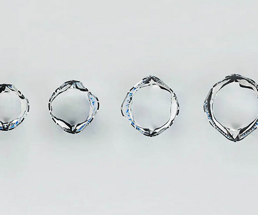
Post-operative expansion is designed to allow for increasing graft diameter, which may enable the graft to match growth in pediatric populations or help to avoid restenosis (narrowing) in adults. Pre-operatively, the controlled dilation may help address unique anatomies. pediatric hospital centers.
Harmony TPV system clinical trials demonstrate ease of implant, conformability to the anatomy, and strong clinical and hemodynamic outcomes for congenital heart disease patients up to three years.
Advances in pediatric cardiac surgery have resulted in a recent growing epidemic of children and young adults with congenital heart diseases (CHDs). In these patients, congenital defects themselves, surgical operations and remaining lesions may alter cardiac anatomy and impact the mechanical performance of both ventricles.
Using machine learning algorithms, InView’s Categorical Hanging Image Protocol(CHIP) automatically organizes images by anatomy and pathology categories to intelligently display or “hang” current and prior studies in a way that significantly improves the efficiency of single or multi-modality image review. As well, by incorporating Us2.ai’s
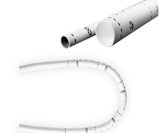
The prototype valve has two leaflets crafted from a polymer material that has a well-established history of being used as a leaflet for pediatric pulmonary valves. To their success, they discovered that this device could be fitted and expanded in sync with the growth of the heart anatomy.
A good knowledge of the anatomy of the heart is needed for interpretation of images from each view. Subcostal view is a favourite view of pediatric echocardiographers. The four common locations at which the echocardiographic transducer is placed for imaging are the parasternal, apical, subcostal, and suprasternal.
BackgroundObesity is associated with diminished myocardial function as measured by strain echocardiography in children and young adults with normal cardiac anatomy. Journal of the American Heart Association, Ahead of Print. Data are lacking about the effect of obesity on myocardial strain in patients with a single ventricle.
For transcatheter ablation in pediatric settings, radiofrequency (RF) ablation was first used in the 1990s, while cryoablation was introduced in 2003 and nowadays represents the most used energy for AVNRT ablation in this population.
We organize all of the trending information in your field so you don't have to. Join thousands of users and stay up to date on the latest articles your peers are reading.
You know about us, now we want to get to know you!
Let's personalize your content
Let's get even more personalized
We recognize your account from another site in our network, please click 'Send Email' below to continue with verifying your account and setting a password.


Let's personalize your content