This site uses cookies to improve your experience. To help us insure we adhere to various privacy regulations, please select your country/region of residence. If you do not select a country, we will assume you are from the United States. Select your Cookie Settings or view our Privacy Policy and Terms of Use.
Cookie Settings
Cookies and similar technologies are used on this website for proper function of the website, for tracking performance analytics and for marketing purposes. We and some of our third-party providers may use cookie data for various purposes. Please review the cookie settings below and choose your preference.
Used for the proper function of the website
Used for monitoring website traffic and interactions
Cookie Settings
Cookies and similar technologies are used on this website for proper function of the website, for tracking performance analytics and for marketing purposes. We and some of our third-party providers may use cookie data for various purposes. Please review the cookie settings below and choose your preference.
Strictly Necessary: Used for the proper function of the website
Performance/Analytics: Used for monitoring website traffic and interactions
All patients were pre-TAVR assessed by transthoracic echocardiography and computed tomography of the aortic valve (AV) and relevant left cardiac and vascular anatomy. Nanjing) to evaluate its safety and efficacy.Methods130 high risk patients with symptomatic severe AS from 11 institutions were treated with the novel Xcor system.
Leadless pacemakers are an alternative to transvenous devices for patients at high risk for lead-related complications. However, conventional implantation is done via femoral access, which may not be possible in cases with variant venous anatomy like interrupted IVC.
Persistent left superior vena cava (PLSVC) is a common anomaly in the thoracic venous system, accounting for 0.2-4.3% of all congenital cardiac anomalies However, isolated PLSVC may occur in 10-20% of PLSVC cases.
ABSTRACT Introduction A leadless pacemaker (LLPM) was recommended for a patient with intermittent complete heart block and near-syncope. Methods and Results Delivery of LLPM is through a large sheath that has limited deflection and steerability. The ventricular LLPM was successfully fixated.
Methods We present the first described case of using two leadless pacing systems manufactured by separate companies implanted within the same patient to provide atrial and ventricular pacing due to complex congenital anatomy. Laser-lead extraction and temporary atrial pacemaker placement was performed.
The challenge of implanting cardiac rhythm devices in patients with complex anatomy has been described in the literature, but cases with extremely abnormal axis are limited.
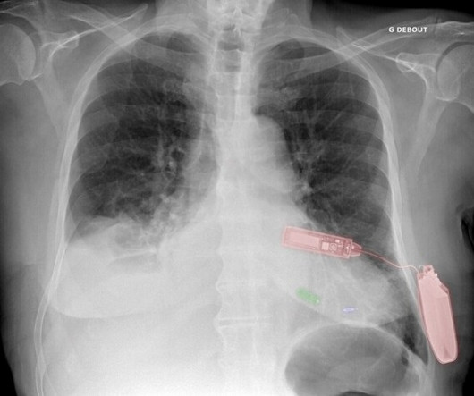
The primary reason is, the LV epicardial lead pacing site was pre-selected by the coronary sinus anatomy. Green: Micra leadless pacemaker; blue: WiSE-CRT system LV endocardial electrode; and red: WiSE-CRT system subcutaneous battery and ultrasound generator. CRT, cardiac resynchronization therapy. 2021 May 21;23(5):740-747.
The primary outcome was mortality, while secondary outcomes included in-hospital complications such as stroke and pacemaker implantation and transcatheter heart valve hemodynamic performance.RESULTS:The number of patients with AS with type 0 bicuspid, type 1 bicuspid, and tricuspid aortic valve anatomy was 328, 302, and 642, respectively.
A temporary pacemaker was implanted, and she was admitted to the ICU with cardiogenic shock. Prompt cath to define the anatomy should be expedited. She was worked as a full code, and ROSC was achieved. She was taken to the cath lab, where she was found to have 100% in-stent restenosis of the proximal LAD. She could not be resuscitated.
His unique cath film demonstration removes all doubt about the anatomy — with the clearest illustration of acute septal perforator occlusion that I have seen! . == MY Comment , by K EN G RAUER, MD ( 3/20 /2024 ): == Superb and enlightening discussion of today's case by Dr. Willy Frick! CLICK HERE — for more on fusion beats.
Rapid Fire Challenging Structural Heart Imaging Cases with Heart Team Panel; Follow-up of Pacemakers and ICDS for the Non-electrophysiologist; The Real Reasons Your Patient with Heart Failure was Readmitted: Noncardiac Comorbidities, Geriatric Syndromes and Social Determinants of Health; and Death by a Thousand Cuts!
To revise the anatomy lessons, this is the external jugular vein and this is the internal jugular vein. Thrombus can sometimes occur when there is a central venous catheter or a multiple pacemaker or defibrillator leads there that can cause thrombus formation. Obstruction could be due to tumour or even a thrombus.
We organize all of the trending information in your field so you don't have to. Join thousands of users and stay up to date on the latest articles your peers are reading.
You know about us, now we want to get to know you!
Let's personalize your content
Let's get even more personalized
We recognize your account from another site in our network, please click 'Send Email' below to continue with verifying your account and setting a password.


Let's personalize your content