Remove Anatomy Remove Ischemia Remove STEMI
article thumbnail

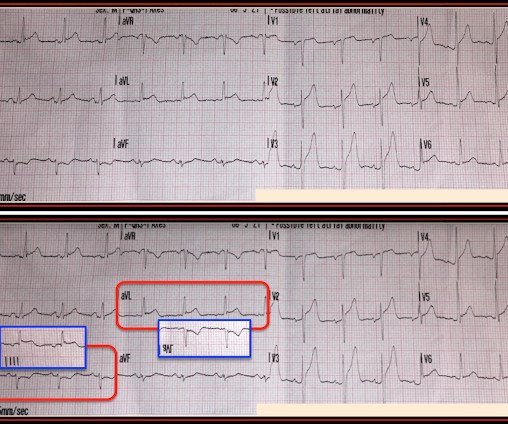
Chest pain and new regional/reciprocal ECG changes compared to previous ECGs: code STEMI?

Dr. Smith's ECG Blog

The admission and discharge diagnosis both attributed the ECG changes and echo findings to ischemia. The biggest problem with STEMI criteria are false negatives – because this costs patient’s myocardium, with greater mortality and morbidity. baseline ECGs may fluctuate over time, and not necessarily represent dynamic ischemia 4.

article thumbnail

90 year old with acute chest and epigastric pain, and diffuse ST depression with reciprocal STE in aVR: activate the cath lab?

Dr. Smith's ECG Blog

This has been termed a “STEMI equivalent” and included in STEMI guidelines, suggesting this patient should receive dual anti-platelets, heparin and immediate cath lab activation–or thrombolysis in centres where cath lab is not available. His response: “subendocardial ischemia.

article thumbnail

5 Cardiologists said this is not a STEMI. But was it an OMI?

Dr. Smith's ECG Blog

Over the next few hours, four other general cardiologists "signed off on the initial ECG without recognizing STEMI." Learning Points: STEMI criteria misses 25-40% of OMI, like this case for example. A millimeter definition of acute STEMI should not be needed to justify the need for prompt cardiac catheterization.

STEMI 52
article thumbnail

A 50-something with chest pain. Is there OMI? And what is the rhythm?

Dr. Smith's ECG Blog

The fact that R waves 2 through 6 are junctional does make ischemia more difficult to interpret -- but not impossible. Back to the assessment of ischemia: Returning to the ECG, the leads that catch my eye first are -- I, II, V4, V5, V6. For national registry purposes, this will be incorrectly classified as a STEMI.) < 0.049).

article thumbnail

ECG Blog #400 — Is this a NSTEMI?

Ken Grauer, MD

Subendocardial Ischemia from another Cause ( ie, sustained tachyarrhythmia; cardiac arrest; shock or profound hypotension; GI bleeding; anemia; "sick patient" , etc. ). To EMPHASIZE: This pattern of diffuse Subendocardial Ischemia does not suggest acute coronary occlusion ( ie, it is not the pattern of an acute MI ).

Blog 100
article thumbnail

ECG Blog #364 — VT in Need of Cardioversion?

Ken Grauer, MD

Looking at the location of ST elevation ( ie, in the inferior leads — and in V4,5,6 ) — and the location of ST depression ( ie, in leads I,aVL; V1,V2 ) — this ECG picture suggests an ongoing acute infero - postero - lateral STEMI. What is S hark F in M orphology ? Cardiopulmonary resuscitation was promptly started — but was unsuccessful.

Blog 78
article thumbnail

ECG Blog #367 — Recognize The KEY Finding?

Ken Grauer, MD

This qualifies as " dynamic " ST-T wave change — and in a patient with new chest pain, this is indication for prompt cath to define the anatomy and ensure reperfusion. ECG Blog #184 — illustrates the "magical" mirror-image opposite relationship with acute ischemia between lead III and lead aVL ( featured in Audio Pearl #2 in this blog post ).

Blog 78