This site uses cookies to improve your experience. To help us insure we adhere to various privacy regulations, please select your country/region of residence. If you do not select a country, we will assume you are from the United States. Select your Cookie Settings or view our Privacy Policy and Terms of Use.
Cookie Settings
Cookies and similar technologies are used on this website for proper function of the website, for tracking performance analytics and for marketing purposes. We and some of our third-party providers may use cookie data for various purposes. Please review the cookie settings below and choose your preference.
Used for the proper function of the website
Used for monitoring website traffic and interactions
Cookie Settings
Cookies and similar technologies are used on this website for proper function of the website, for tracking performance analytics and for marketing purposes. We and some of our third-party providers may use cookie data for various purposes. Please review the cookie settings below and choose your preference.
Strictly Necessary: Used for the proper function of the website
Performance/Analytics: Used for monitoring website traffic and interactions
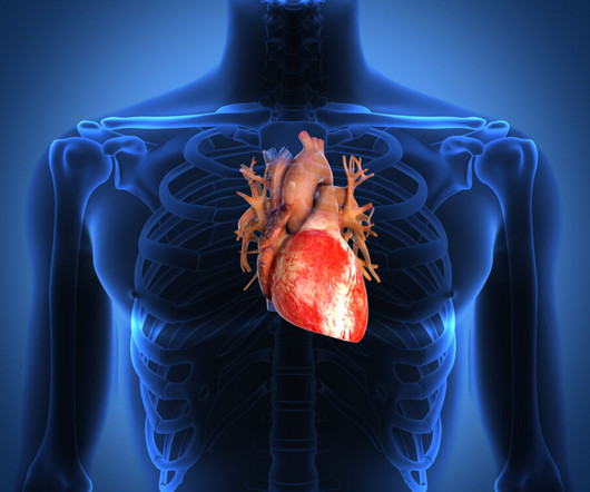
The post Anatomy of the Mitral Valve appeared first on All About Cardiovascular System and Disorders. The mitral valve complex consists of the mitral annulus, mitral valve leaflets, the chordae tendineae and the papillary muscles. The mitral leaflets are the anterior and posterior leaflets.
Nuclear Cardiology See if you’re ready for the Nuclear Cardiology boards by answering these hard-hitting sample questions plucked from our very own nuclear cardiology question bank. A 48 year-old female with hypertension, hyperlipidemia, chronic low back pain, and bilateral lower extremity neuropathy.
That is, pulmonary artery is transposed over to the right ventricle, and aorta over to the left ventricle, so that normal anatomy is restored. In simple terms, arterial switch is restoring the normal connections. But that is easily said than done. It is much more difficult than you think from this simple block diagram.
To revise the anatomy lessons, this is the external jugular vein and this is the internal jugular vein. Transcript of the video: Now we will discuss the basic principles of evaluation of jugular venous pressure and jugular venous pulse. These are assessed in the internal jugular vein and not in the external jugular vein.
Evaluation of diastolic characteristics of LV and LV and coronary anatomy evaluation are other diagnostic uses of cath in HCM. Degree of outflow obstruction can be documented along with the classical Brockenbrough-Braunwald-Morrow sign. The sign is an increased LVOT gradient after a ventricular premature complex.
A good knowledge of the anatomy of the heart is needed for interpretation of images from each view. The four common locations at which the echocardiographic transducer is placed for imaging are the parasternal, apical, subcostal, and suprasternal. Parasternal views are often obtained first, followed by apical, subcostal, and suprasternal.
We organize all of the trending information in your field so you don't have to. Join thousands of users and stay up to date on the latest articles your peers are reading.
You know about us, now we want to get to know you!
Let's personalize your content
Let's get even more personalized
We recognize your account from another site in our network, please click 'Send Email' below to continue with verifying your account and setting a password.


Let's personalize your content