This site uses cookies to improve your experience. To help us insure we adhere to various privacy regulations, please select your country/region of residence. If you do not select a country, we will assume you are from the United States. Select your Cookie Settings or view our Privacy Policy and Terms of Use.
Cookie Settings
Cookies and similar technologies are used on this website for proper function of the website, for tracking performance analytics and for marketing purposes. We and some of our third-party providers may use cookie data for various purposes. Please review the cookie settings below and choose your preference.
Used for the proper function of the website
Used for monitoring website traffic and interactions
Cookie Settings
Cookies and similar technologies are used on this website for proper function of the website, for tracking performance analytics and for marketing purposes. We and some of our third-party providers may use cookie data for various purposes. Please review the cookie settings below and choose your preference.
Strictly Necessary: Used for the proper function of the website
Performance/Analytics: Used for monitoring website traffic and interactions
Echocardiogram An echocardiogram uses sound waves to produce a detailed image of the heart, allowing doctors to see the size of the heart chambers and how well the heart is pumping blood. The following diagnostic tools are commonly used: 1.
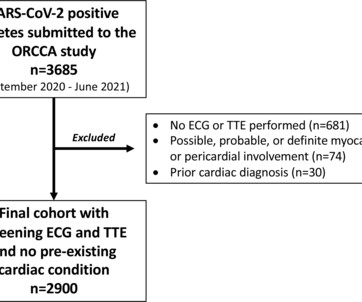
Athletes with an ECG and transthoracic echocardiogram (TTE) and no pre-existing conditions were included. Overall, 6 (0.2%) athletes had major conditions; however, coronary anatomy and aortic dimensions were inconsistently reported and pathology may have been missed.
At 30 days, 11 of 12 surviving patients had an available echocardiogram; mitral regurgitation severity was trace/none in 90.9% (10/11) and mild in 9.1% (1/11).CONCLUSIONS:The CONCLUSIONS:The AltaValve system shows promising early procedural and clinical results for the unique anatomy of patients with atrial functional mitral regurgitation.
Diagnostic cardiac catheterization may be needed especially in tetralogy of Fallot with pulmonary atresia, to assess the pulmonary anatomy, including size and distribution of peripheral pulmonary arteries. Another important role is for detection of coronary anomalies, which can also be seen on echocardiogram sometimes.
To their success, they discovered that this device could be fitted and expanded in sync with the growth of the heart anatomy. Before implantation, doctors can adjust the valve diameter to match the patient’s heart anatomy.
Methods We present the first described case of using two leadless pacing systems manufactured by separate companies implanted within the same patient to provide atrial and ventricular pacing due to complex congenital anatomy. Laser-lead extraction and temporary atrial pacemaker placement was performed.
Although he had a normal echocardiogram and stress test a year ago at a different hospital, due to his symptoms and intermediate-high risk probability of coronary artery disease (CAD), the decision was made to proceed with a cardiac catheterization using a trans-radial approach with a horizontal sweep technique.
Using machine learning algorithms, InView’s Categorical Hanging Image Protocol(CHIP) automatically organizes images by anatomy and pathology categories to intelligently display or “hang” current and prior studies in a way that significantly improves the efficiency of single or multi-modality image review. As well, by incorporating Us2.ai’s
The image shown here is an animated 2 dimensional echocardiogram. This one is an older mode known as time-motion mode or M-Mode echocardiogram. A good knowledge of the anatomy of the heart is needed for interpretation of images from each view. Unlike the previous 2 dimensional imaging, this is a single dimensional imaging.
The best course is to wait until the anatomy is defined by angio, then if proceeding to PCI, add Cangrelor (an IV P2Y12 inhibitor) I sent the ECG and clinical information of a 90-year old with chest pain to Dr. McLaren. See this case: what do you think the echocardiogram shows in this case?
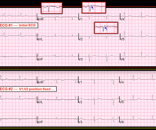
She had a normal echocardiogram, with normal shortening and thickening of the septum. Variation in body habitus and chest wall anatomy may also sometimes account for unexpected ECG findings. American Journal of Emergency Medicine 36(5):865-870; May 2018. Here are other cases with lead misplacement: [link] Learning Points: 1.
Echocardiogram showing thickened interventricular septum and mitral regurgitation in HCM. Evaluation of diastolic characteristics of LV and LV and coronary anatomy evaluation are other diagnostic uses of cath in HCM. SAM in HCM Systolic anterior movement of mitral valve occurs in 30 – 60%, but it is not specific.
Compare to the anatomy after stenting: The lower of the 2 now easily seen branches is the circumflex, now with excellent flow. Next day echocardiogram showed inferolateral hypokinesia with an EF of %45-50. This is seen just millimeters beyond the tip of the catheter. The patient recovered well. His peak troponin was over 5000 ng/L.
First in slow motion with a freeze frame with annotated vessel anatomy, then at normal speed. Echocardiogram showed inferior hypokinesis. Angiogram is shown below. As always I use the same color conventions for vessels as the rest of my angiography guide. Troponin was rising when last checked, 8928 ng/L.
Right Ventricular Outflow Tract (RVOT) Tachycardia Fascicular Tachycardia Bundle branch re-entry tachycardia Discussion : The diagnosis and management of the i diopathic VTs a re predicated on an understanding of the mechanism, relevant cardiac anatomy, and associated ECG signatures.
We organize all of the trending information in your field so you don't have to. Join thousands of users and stay up to date on the latest articles your peers are reading.
You know about us, now we want to get to know you!
Let's personalize your content
Let's get even more personalized
We recognize your account from another site in our network, please click 'Send Email' below to continue with verifying your account and setting a password.


Let's personalize your content