This site uses cookies to improve your experience. To help us insure we adhere to various privacy regulations, please select your country/region of residence. If you do not select a country, we will assume you are from the United States. Select your Cookie Settings or view our Privacy Policy and Terms of Use.
Cookie Settings
Cookies and similar technologies are used on this website for proper function of the website, for tracking performance analytics and for marketing purposes. We and some of our third-party providers may use cookie data for various purposes. Please review the cookie settings below and choose your preference.
Used for the proper function of the website
Used for monitoring website traffic and interactions
Cookie Settings
Cookies and similar technologies are used on this website for proper function of the website, for tracking performance analytics and for marketing purposes. We and some of our third-party providers may use cookie data for various purposes. Please review the cookie settings below and choose your preference.
Strictly Necessary: Used for the proper function of the website
Performance/Analytics: Used for monitoring website traffic and interactions
Echocardiogram An echocardiogram uses sound waves to produce a detailed image of the heart, allowing doctors to see the size of the heart chambers and how well the heart is pumping blood. Regular physical activity can strengthen the heart and improve circulation. The following diagnostic tools are commonly used: 1.
Circulation: Cardiovascular Interventions, Ahead of Print. At 30 days, 11 of 12 surviving patients had an available echocardiogram; mitral regurgitation severity was trace/none in 90.9% (10/11) and mild in 9.1% (1/11).CONCLUSIONS:The Class III/IV New York Heart Association status was reduced from 79% at baseline to 0% at 30 days.
To their success, they discovered that this device could be fitted and expanded in sync with the growth of the heart anatomy. Before implantation, doctors can adjust the valve diameter to match the patient’s heart anatomy.
Circulation, Volume 150, Issue Suppl_1 , Page A4140682-A4140682, November 12, 2024. Introduction:Dextrocardia is a rare congenital condition where the heart's apex points to the right, with an incidence of about 0.01%. Patients usually have a normal life expectancy unless other structural heart diseases are present.
Compare to the anatomy after stenting: The lower of the 2 now easily seen branches is the circumflex, now with excellent flow. Next day echocardiogram showed inferolateral hypokinesia with an EF of %45-50. Circulation 2002; 105(4): 539-42. This is seen just millimeters beyond the tip of the catheter. The patient recovered well.
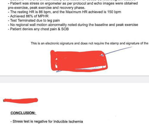
The shrewd cardiologist did a bicycle ergometry and simultaneous echocardiogram without any drugs or injections. We are obsessed with anatomy. Our flawed intellect keeps asking this question: How can I trust physiology (Flow) without documenting a good anatomy? A good epicardial anatomy rarely guarantee good physiology. (It
Right Ventricular Outflow Tract (RVOT) Tachycardia Fascicular Tachycardia Bundle branch re-entry tachycardia Discussion : The diagnosis and management of the i diopathic VTs a re predicated on an understanding of the mechanism, relevant cardiac anatomy, and associated ECG signatures. Nossen evaluation included: A normal Echo.
We organize all of the trending information in your field so you don't have to. Join thousands of users and stay up to date on the latest articles your peers are reading.
You know about us, now we want to get to know you!
Let's personalize your content
Let's get even more personalized
We recognize your account from another site in our network, please click 'Send Email' below to continue with verifying your account and setting a password.


Let's personalize your content