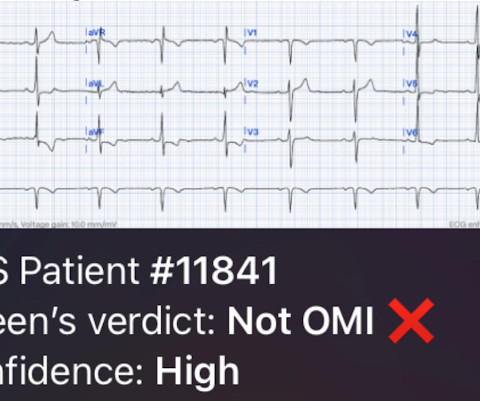
Remove Anatomy Remove Chest Pain Remove Ischemia
article thumbnail

Chest pain and new regional/reciprocal ECG changes compared to previous ECGs: code STEMI?

Dr. Smith's ECG Blog

Written by Jesse McLaren A 45 year old presented with two weeks of recurring non-exertional chest pain, now constant for an hour. Because of the ECG changes in a patient with chest pain, and with inferolateral hypokinesis on POCUS, the cath lab was activated. Below is old and then new ECG (old on top; new below).

article thumbnail

A 50-something with chest pain. Is there OMI? And what is the rhythm?

Dr. Smith's ECG Blog

Written by Willy Frick A man in his 50s with history of hypertension, hyperlipidemia, and a 30 pack-year smoking history presented to the ER with 1 hour of acute onset, severe chest pain and diaphoresis. The fact that R waves 2 through 6 are junctional does make ischemia more difficult to interpret -- but not impossible.

article thumbnail

A teenager with chest pain, a troponin below the limit of detection, and "benign early repolarization"

Dr. Smith's ECG Blog

No prior exertional complaints of chest pain, dizziness, lightheadedness, or undue shortness of breath. He denied headache or neck pain associated with exertion. I sent this ECG to Dr. Smith, with the only information that it is a 17 year old with chest pain. 24 yo woman with chest pain: Is this STEMI?

article thumbnail

Chest pain, and Cardiology didn't take the hint from the ICD

Dr. Smith's ECG Blog

Submitted and written by Megan Lieb, DO with edits by Bracey, Smith, Meyers, and Grauer A 50-ish year old man with ICD presented to the emergency department with substernal chest pain for 3 hours prior to arrival. At this time he reported ongoing chest pain and was given aspirin and nitroglycerin. J Am Heart Assoc.

article thumbnail

ECG Blog #400 — Is this a NSTEMI?

Ken Grauer, MD

Subendocardial Ischemia from another Cause ( ie, sustained tachyarrhythmia; cardiac arrest; shock or profound hypotension; GI bleeding; anemia; "sick patient" , etc. ). To EMPHASIZE: This pattern of diffuse Subendocardial Ischemia does not suggest acute coronary occlusion ( ie, it is not the pattern of an acute MI ).

Blog 100
article thumbnail

ECG Blog #367 — Recognize The KEY Finding?

Ken Grauer, MD

But, in a patient who presents to the ED for new chest pain — seeing these subtle findings that are localized to leads V2- thru -V4 should at the least make you consider acute posterior OMI ( O cclusion-based MI ) — until you prove otherwise. To EMPHASIZE: These are subtle findings. What do YOU think?

Blog 78
article thumbnail

90 year old with acute chest and epigastric pain, and diffuse ST depression with reciprocal STE in aVR: activate the cath lab?

Dr. Smith's ECG Blog

The best course is to wait until the anatomy is defined by angio, then if proceeding to PCI, add Cangrelor (an IV P2Y12 inhibitor) I sent the ECG and clinical information of a 90-year old with chest pain to Dr. McLaren. His response: “subendocardial ischemia. The patient had mild but diffuse abdominal tenderness.