This site uses cookies to improve your experience. To help us insure we adhere to various privacy regulations, please select your country/region of residence. If you do not select a country, we will assume you are from the United States. Select your Cookie Settings or view our Privacy Policy and Terms of Use.
Cookie Settings
Cookies and similar technologies are used on this website for proper function of the website, for tracking performance analytics and for marketing purposes. We and some of our third-party providers may use cookie data for various purposes. Please review the cookie settings below and choose your preference.
Used for the proper function of the website
Used for monitoring website traffic and interactions
Cookie Settings
Cookies and similar technologies are used on this website for proper function of the website, for tracking performance analytics and for marketing purposes. We and some of our third-party providers may use cookie data for various purposes. Please review the cookie settings below and choose your preference.
Strictly Necessary: Used for the proper function of the website
Performance/Analytics: Used for monitoring website traffic and interactions
In an era of rapidly expanding use of transcatheter aortic valve implantation (TAVI), the management of patients with bicuspid aortic valve (BAV) disease is far less well established than in those with trileaflet anatomy.
The mitral valve complex consists of the mitral annulus, mitral valve leaflets, the chordae tendineae and the papillary muscles. The mitral leaflets are the anterior and posterior leaflets. The post Anatomy of the Mitral Valve appeared first on All About Cardiovascular System and Disorders.
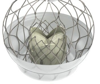
Food and Drug Administratio n ( FDA ) for the AltaValve System , a transcatheter mitral valve replacement (TMVR) device. MR occurs when blood flows backward through the mitral valve and into the atrium each time the left ventricle contracts. milla1cf Wed, 05/08/2024 - 11:02 May 8, 2024 — 4C Medical Technologies, Inc. ("4C
All patients were pre-TAVR assessed by transthoracic echocardiography and computed tomography of the aortic valve (AV) and relevant left cardiac and vascular anatomy. Nanjing) to evaluate its safety and efficacy.Methods130 high risk patients with symptomatic severe AS from 11 institutions were treated with the novel Xcor system.
(MedPage Today) -- PARIS -- Patients with questionable suitability for transcatheter aortic valve replacement (TAVR) in fact fared worse in the short term after the catheter-based procedure than after surgery, the NOTION-2 randomized trial showed.
BACKGROUND:Data concerning the outcomes of transcatheter aortic valve replacement in type 0 bicuspid aortic stenosis (AS) are scarce. Ascending aortic diameter was the single predictor of 1-year mortality in type 0 bicuspid patients (hazard ratio, 1.59 [95% CI, 1.03–2.44];P=0.035). Poverall=0.522; 1 year: 10% versus 2.3%
Our recent Mitral Valve Training, held on January 10-11, emerged as a beacon of excellence, bringing together cardiologists from across the globe for an immersive experience. Concurrently, our anatomy session, featuring bull hearts and anatomy tools, facilitated a deeper understanding of cardiac anatomy.
Background There are different types of transcatheter mitral valve repair (TMVr) currently in clinical use, including leaflet approximation, annular cinching, and restoration of the chordal apparatus of the mitral valve (MV). Aims To evaluate the procedural and clinical outcome of COMBO therapies compared with M-TEER alone.
milla1cf Mon, 01/08/2024 - 14:35 January 8, 2024 — University Hospitals (UH) Harrington Heart & Vascular Institute recently became the first center in the world to implant Medtronic’s Penditure Left Atrial Appendage (LAA) Exclusion System through a minimally invasive approach during a mitral valve repair procedure.
BACKGROUND:Many patients with atrial functional mitral regurgitation are not suitable candidates for surgery or transcatheter repair. Technical success and mitral regurgitation reduction from severe to none/trace were achieved in all cases. There were no cases of left ventricular outflow tract obstruction.
ADN CoE recently held an exceptional two-day training focused on the latest techniques for treating mitral valve disease. Ismail Ateş and Dr. Zeynettin Kaya; renowned experts in mitral valve interventions, this course offered an amazing opportunity for continued education. Led by Assoc. stay tuned for what's coming next.
Our recent Mitral Valve Training, held on January 10-11, emerged as a beacon of excellence, bringing together cardiologists from across the globe for an immersive experience. Concurrently, our anatomy session, featuring bull hearts and anatomy tools, facilitated a deeper understanding of cardiac anatomy.
Focal AT at the aorta-mitral annulus conjunction (AMC) is uncommon. Hence, the electrophysiological and ablation target characteristics are poorly described.
Focusing on mitral and tricuspid valve diseases , Capstans treatment combines transcatheter implantation of a folded valve replacement with its X-ray and ultrasound-guided robot to align the low-profile implant with the beating heart valve. The post Capstans Robotic MR/TR Frontier Funding appeared first on Cardiac Wire.
BackgroundWe previously reported procedural and 30‐day outcomes of a German early multicenter experience with the PASCAL system for severe mitral regurgitation (MR). Journal of the American Heart Association, Ahead of Print.
The MitraClip procedure, is designed to reduce mitral regurgitation (MR) by approximating the mitral valve leaflets, can alter the direction or nature of residual MR, including potentially converting a central MR jet into an eccentric one. This could redirect the residual regurgitant flow. Implication of new onset eccentric jet 1.Eccentric
TriClip leverages the same clip-based technology as Abbott's leading MitraClip device – which has treated more than 200,000 people with leaky mitral valves (mitral regurgitation) – but was specifically designed to treat the tricuspid valve's complex anatomy.
Leon, Standardized Definitions for Bioprosthetic Valve Dysfunction Following Aortic or Mitral Valve Replacement: JACC State-of-the-Art Review, Journal of the American College of Cardiology, Volume 80, Issue 5, 2022, Pages 545-561, ISSN 0735-1097, [link]. Grayburn, Patrizio Lancellotti, Vinod H. Thourani, Jeroen J. Bax, Michael J.
In the process, improving the ejection fraction and possibly reducing mitral regurgitation. The primary reason is, the LV epicardial lead pacing site was pre-selected by the coronary sinus anatomy. Still, the optimal benefit of CRT concept has been difficult to extract from this device.
The bicuspid or mitral valve is the left atrioventricular valve is. These valves consist of tissue that is about as thick as a piece of paper. Like the heart chambers, there are four heart valves between each of the chambers. The tricuspid valve is the right atrioventricular valve.
The post ectopic increase in the murmur is a hallmark of hypertrophic obstructive cardiomyopathy, which differentiates it clinically from mitral valve prolapse. Echocardiography in HCM Important echocardiographic features include mitral regurgitation and left ventricular outflow tract obstruction.
Tracing in the lower part is tissue Doppler imaging from the medial mitral annulus. A good knowledge of the anatomy of the heart is needed for interpretation of images from each view. Opening and closing movements of the aortic and mitral valves are visible. The images shown so far were from transthoracic echocardiography.
Compare to the anatomy after stenting: The lower of the 2 now easily seen branches is the circumflex, now with excellent flow. For example, mid-anterolateral and mid-inferior segments generally harbor papillary muscles and infarction of these segments may result in acute mitral regurgitation due to papillary muscle dysfunction or rupture.
It needs a good knowledge of anatomy, physiology of inter & Intra valvular hemodynamics.It It needs a good knowledge of anatomy, physiology of inter & Intra valvular hemodynamics.It Mind you, even an innocuous episode of fever, associated dyspnea, and tachycardia can elevate the mitral gradient and sound a false alarm.
ABSTRACT Background Achieving a durable mitral line block using radiofrequency as a part of an anatomical approach for ablation in patients with persistent atrial fibrillation or for treating peri-mitral flutter has always been challenging due to the complex anatomy of the mitral isthmus.
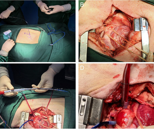
Surgical techniques included mitral valve replacement, mitral valve repair, aortic valve replacement, OZAKI procedure, ascending aorta replacement, and left ventricular assist device implantation. All animals underwent cardiac valve surgeries via left thoracotomy with CPB.
We organize all of the trending information in your field so you don't have to. Join thousands of users and stay up to date on the latest articles your peers are reading.
You know about us, now we want to get to know you!
Let's personalize your content
Let's get even more personalized
We recognize your account from another site in our network, please click 'Send Email' below to continue with verifying your account and setting a password.


Let's personalize your content