This site uses cookies to improve your experience. To help us insure we adhere to various privacy regulations, please select your country/region of residence. If you do not select a country, we will assume you are from the United States. Select your Cookie Settings or view our Privacy Policy and Terms of Use.
Cookie Settings
Cookies and similar technologies are used on this website for proper function of the website, for tracking performance analytics and for marketing purposes. We and some of our third-party providers may use cookie data for various purposes. Please review the cookie settings below and choose your preference.
Used for the proper function of the website
Used for monitoring website traffic and interactions
Cookie Settings
Cookies and similar technologies are used on this website for proper function of the website, for tracking performance analytics and for marketing purposes. We and some of our third-party providers may use cookie data for various purposes. Please review the cookie settings below and choose your preference.
Strictly Necessary: Used for the proper function of the website
Performance/Analytics: Used for monitoring website traffic and interactions
Patient is pain free and clearly has Wellens' syndrome: 1) pain free episode following an episode of angina, typical Pattern A (biphasic, terminal T-wave inversion with an initial upsloping ST Segment) findings, preserved R-waves. Angiography : --Culprit for the patient's unstable angina/Wellen syndrome is a ruptured plaque in the mid LAD. --As
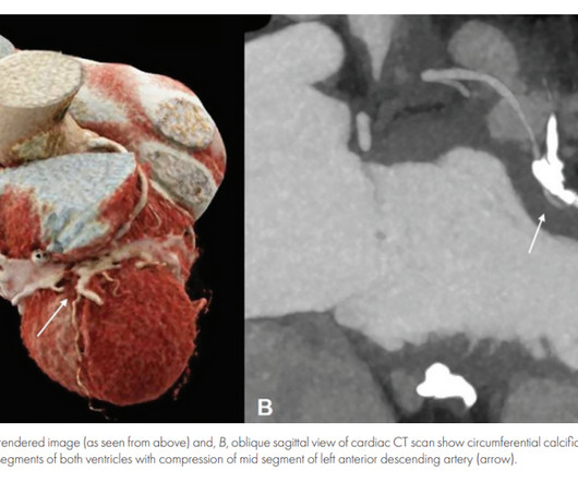
If you understand the pericardial anatomy fully, you can call yourself real master of clinical anatomy. There has been many reports of patients with angina in CP (Ref 1). Angina caused by calcific constrictive pericarditis. Structure and Anatomy of the Human Pericardium. Fortunately, it escapes in many.
In addition, the criteria require the absence of precordial Q waves, the presence of history of angina, and normal or slightly elevated cardiac serum markers. Wellens is a glorified subset of ACS. It can be referred to as an ACS in a confused state of evolution. Most often a critical mechanical LAD lesion is noted. Reference 1.
Coronary anatomy and SYNTAX(Synergy between percutaneous coronary intervention with Taxus and cardiac surgery) scores were measured using coronary computed tomography angiography. Diabetes was present in 28% and multivessel disease in 51%. Most clinical scenarios favored invasive for better 1-year health status.
We present the cumulative percutaneous coronary intervention (PCI) data of all comers (stable angina and acute coronary syndromes [ACS]) who presented to Hadi Clinic between January 2018 and December 2020. Pre-procedural data included patients’ baseline characteristics (age, gender, clinical presentation and comorbidities).
Patients with dextrocardia present a diagnostic challenge, particularly in the context of acute coronary syndrome.Case Presentation:A 49-year-old male with a medical history of dextrocardia, hypothyroidism, dyslipidemia and hypertension was referred to a cardiologist by his primary physician due to a 3-week history of unstable angina.
For example, if a coronary artery becomes blocked due to plaque buildup (a condition known as coronary artery disease), the heart muscle may not receive enough oxygen, leading to chest pain (angina) or, in more severe cases, a heart attack. CAD is one of the leading causes of heart attacks.
Session 510) To Treat or Not to Treat Anatomy and Ischemia? (Session 508) Battle of the Imagers - Jeopardy Edition! Session 509) Who Wants to Be a Millionaire in Eradicating Vascular Medicine Disparities?
Angina is another common symptom due the hypertrophy which causes a coronary supply demand mismatch Symptoms of HCM include syncope/near syncope, which could be precipitated by exertion, hypovolemia, rapid standing, Valsalva manoeuvre, diuretics, vasodilators or arrhythmia. The role of cath now a days is mostly for septal ablation.
One big chunk of ACS-UA is secondary UA where there is increased demand as in stable angina with tachycardia*. In these patients there is no plaque triggered ACS. For example, in a febrile patient who has associated HT, anemia, etc., You can’t err at the same time , you are not supposed to treat inappropriate as well.
First in slow motion with a freeze frame with annotated vessel anatomy, then at normal speed. Even though guidelines say that patients with high-risk features, refractory angina, instability, etc. Angiogram is shown below. As always I use the same color conventions for vessels as the rest of my angiography guide.
We organize all of the trending information in your field so you don't have to. Join thousands of users and stay up to date on the latest articles your peers are reading.
You know about us, now we want to get to know you!
Let's personalize your content
Let's get even more personalized
We recognize your account from another site in our network, please click 'Send Email' below to continue with verifying your account and setting a password.


Let's personalize your content