This site uses cookies to improve your experience. To help us insure we adhere to various privacy regulations, please select your country/region of residence. If you do not select a country, we will assume you are from the United States. Select your Cookie Settings or view our Privacy Policy and Terms of Use.
Cookie Settings
Cookies and similar technologies are used on this website for proper function of the website, for tracking performance analytics and for marketing purposes. We and some of our third-party providers may use cookie data for various purposes. Please review the cookie settings below and choose your preference.
Used for the proper function of the website
Used for monitoring website traffic and interactions
Cookie Settings
Cookies and similar technologies are used on this website for proper function of the website, for tracking performance analytics and for marketing purposes. We and some of our third-party providers may use cookie data for various purposes. Please review the cookie settings below and choose your preference.
Strictly Necessary: Used for the proper function of the website
Performance/Analytics: Used for monitoring website traffic and interactions
Furthermore, it aids in planning and conducting safe aortic intervention and assists in deciding on single- or two-staged stent graft procedures. It underscores the value of preoperative CT councils and provides crucial information for interpreting the results.
IntroductionBasilar‐tip aneurysm (BTA) is the most common aneurysm found in the posterior circulation, representing 5–8% of total intracranial aneurysms. For ruptured aneurysms, Adjuvant therapy (BAC or SAC) was used to treat larger dimension aneurysms compared to CE (p = 0.046). vs. 10.7%).
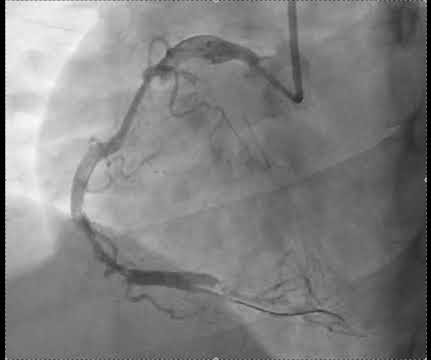
They found an acute lesion of the LAD at the site of the prior stents, including 70% proximal LAD lesion and 95% mid-LAD stenosis with TIMI 3 flow at the time of cath. The LAD lesion was acute and required 3 stents to restore flow. because if it does, then urgent cath to define the anatomy is clearly indicated. Similar findings.
Compare to the anatomy after stenting: The lower of the 2 now easily seen branches is the circumflex, now with excellent flow. Post-myocardial infarction (MI) ventricular septal defects are frequently seen in mid-anteroseptal and apical septal segments, whereas apex and the basal inferior segment are prone to aneurysm formation.
Endovascular aneurysm repair (EVAR) and thoracic endovascular aortic repair (TEVAR) are minimally invasive procedures to treat abdominal, and many thoracic, aortic aneurysms. An aneurysm, an abnormal bulge or ballooning in the wall of a blood vessel, can burst which causes bleeding inside the body and often leads to death. [1]
The report describes heavy plaque in the proximal RCA by IVUS, but no lesions in the previously occluded RPL branch and no stent was deployed. It is consistent with an inferior LV aneurysm. Her ECG afterward is shown below: ECG from five days prior Smith : this shows an old inferior MI with persistent ST elevation.
His initial high sensitivity troponin I returned at 1300 ng/L and given that his cardiac workup was otherwise unremarkable, a CT was obtained to evaluate for pulmonary embolism and aortic aneurysm or dissection but this too was unrevealing. Another EKG was also obtained. ECG at time 82 minutes: What do you think?
We organize all of the trending information in your field so you don't have to. Join thousands of users and stay up to date on the latest articles your peers are reading.
You know about us, now we want to get to know you!
Let's personalize your content
Let's get even more personalized
We recognize your account from another site in our network, please click 'Send Email' below to continue with verifying your account and setting a password.


Let's personalize your content