Remove AFIB Remove Echocardiogram Remove Hospital
article thumbnail

InterAtrial Block

EMS 12-Lead

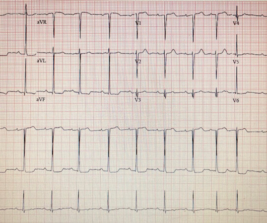
In the pre-hospital setting the varying modalities needed to rule-in/rule-out these causative factors are not available (eg, Chest X-ray, Echocardiogram, etc). And since common things are common, the statistical probability favors Atrial Fibrillation (AFib) as the culprit. AFib can come and go. But the ECG is Sinus!

AFIB 59
article thumbnail

FDA Clears First AI to Aid Heart Failure Detection During Routine Check-ups

DAIC

As a result, many heart failure cases go undiagnosed until symptoms force a specialist or emergency hospital visit, leading to worse patient outcomes and exacerbated healthcare costs.2 Survival and health economic outcomes in heart failure diagnosed at hospital admission versus community settings: a propensity-matched analysis.

article thumbnail

News from EHRA 2024: International Experts Agree on Standards for Catheter Ablation of Atrial Fibrillation

DAIC

AFib Facts and Impacts Atrial fibrillation is the most common cardiac arrhythmia, affecting 2% of individuals worldwide. Before the procedure, patients should have an electrocardiogram (ECG) and echocardiogram (ultrasound of the heart) to check the heart’s rhythm and function.

article thumbnail

A teenager involved in a motor vehicle collision with abnormal ECG

Dr. Smith's ECG Blog

Hopefully a repeat echocardiogram will be performed outpatient. Between 81-95% of life-threatening ventricular dysrhythmias and acute cardiac failure occur within 24-48 hours of hospitalization. Other Arrhythmias ( PACs, PVCs, AFib, Bradycardia and AV conduction disorders — potentially lethal VT/VFib ). No cardiac MRI was done.

article thumbnail

Cryptogenic Stroke

Dr. Sanjay Gupta

Conditions such as AFib are often said to be associated with stroke as a consequence of cardioembolism. All patients should have a 12 lead ECG and be put on a 24 hour heart monitor whilst in hospital. Another way of imaging the heart is via a transesophageal echocardiogram. This is called cardioembolism.

Strokes 52
article thumbnail

90 year old with acute chest and epigastric pain, and diffuse ST depression with reciprocal STE in aVR: activate the cath lab?

Dr. Smith's ECG Blog

See this case: what do you think the echocardiogram shows in this case? With the history of Afib, CTA abdomen was ordered to r/o mesenteric ischemia vs ischemic colitis vs small bowel obstruction. However, STE-aVR with multilead ST depression was associated with 31% in-hospital mortality compared with only 6.2%

article thumbnail

ECG Blog #412 — Is Cardiac Cath Indicated?

Ken Grauer, MD

That said — distinction between "classic" HCM vs the apical HCM for m may be useful because: i ) ECG findings tend to be different ( Lyon et al — Europace 20:102-112iii, 2018 ) ; — ii ) Echo appearance is different when hypertrophy localizes to the apex; and , iii ) There is a significantly greater incidence of AFib with apical HCM.

Blog 148