This site uses cookies to improve your experience. To help us insure we adhere to various privacy regulations, please select your country/region of residence. If you do not select a country, we will assume you are from the United States. Select your Cookie Settings or view our Privacy Policy and Terms of Use.
Cookie Settings
Cookies and similar technologies are used on this website for proper function of the website, for tracking performance analytics and for marketing purposes. We and some of our third-party providers may use cookie data for various purposes. Please review the cookie settings below and choose your preference.
Used for the proper function of the website
Used for monitoring website traffic and interactions
Cookie Settings
Cookies and similar technologies are used on this website for proper function of the website, for tracking performance analytics and for marketing purposes. We and some of our third-party providers may use cookie data for various purposes. Please review the cookie settings below and choose your preference.
Strictly Necessary: Used for the proper function of the website
Performance/Analytics: Used for monitoring website traffic and interactions
I went to the patient's chart: Elderly woman with stuttering chestpain and SOB, and dizziness. This is a very typical ECG for Hypertrophic Cardiomyopathy. Tall R wave in lead V1 and/or early transition in the chest leads ( reflecting increased "septal" forces ). WPW Cardiac arrhythmias ( including AFib ).
The patient has acute chestpain. Tall R wave in lead V1 and/or early transition in the chest leads ( reflecting increased "septal" forces ). WPW Cardiac arrhythmias ( especially AFib ). This was texted to me in real time. What do you think? Here was my answer: "Not ischemia. Maybe HOCM or another form of LVH.
On the other hand — the ST elevation seen in lead V1 is perfectly consistent with LVH and LV "strain" ( ie, The shape of this ST-T wave in lead V1, in association with the deep S wave in this lead — is a mirror-image opposite picture of the typical expected appearance of LVH with "strain" in a lateral chest lead ).
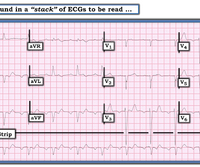
The patient also has a history of AFib and HFmrEF ( = H eart F ailure with M inimally- R educed E jection F raction ). This patient presented to the ED “after a couple of days of chest discomfort”. For clarity in Figure-1 — I have reproduced and labeled this patient’s initial ECG. Why was it Wrong to Think the Rhythm was AFlutter?
Instead, he complained of left chest "itchiness". He had a h/o ischemic cardiomyopathy and right MCA stroke. 9 Hours of ChestPain and Deep Q-waves: Is it too late for Thrombolytics? As per Dr. Smith — this suggests that despite QRS widening, the rhythm in ECG #3 is AFib with a rapid ventricular response.
There was some dyspnea but no chestpain. Tall R wave in lead V1 and/or early transition in the chest leads ( reflecting increased "septal" forces ). WPW Cardiac arrhythmias ( especially AFib ). A young man presented with continuous prolonged generalized weakness, lightheadedness, and presyncope. Here is his ECG.
There was no chestpain — and all troponins were negative. Atrial arrhythmias ( especially AFib or AFlutter ). Smith immediately knew he needed to find out what was going on with this patient! It turned out the patient had cardiac amyloidosis. The presenting complaint was cough and fever from mild Covid pneumonia.
This middle-aged man with no cardiac history but with significant history of methamphetamin and alcohol use presented with chestpain and SOB, worsening over days, with orthopnea. The absence of any wall motion abnormality makes ischemic cardiomyopathy very unlikely. BP:143/99, Pulse 109, Temp 37.2 °C C (99 °F), Resp (!)
We organize all of the trending information in your field so you don't have to. Join thousands of users and stay up to date on the latest articles your peers are reading.
You know about us, now we want to get to know you!
Let's personalize your content
Let's get even more personalized
We recognize your account from another site in our network, please click 'Send Email' below to continue with verifying your account and setting a password.


Let's personalize your content