This site uses cookies to improve your experience. To help us insure we adhere to various privacy regulations, please select your country/region of residence. If you do not select a country, we will assume you are from the United States. Select your Cookie Settings or view our Privacy Policy and Terms of Use.
Cookie Settings
Cookies and similar technologies are used on this website for proper function of the website, for tracking performance analytics and for marketing purposes. We and some of our third-party providers may use cookie data for various purposes. Please review the cookie settings below and choose your preference.
Used for the proper function of the website
Used for monitoring website traffic and interactions
Cookie Settings
Cookies and similar technologies are used on this website for proper function of the website, for tracking performance analytics and for marketing purposes. We and some of our third-party providers may use cookie data for various purposes. Please review the cookie settings below and choose your preference.
Strictly Necessary: Used for the proper function of the website
Performance/Analytics: Used for monitoring website traffic and interactions
Cath lab declined as it is not a STEMI." And now this finding is even formally endorsed as a "STEMI equivalent" in the 2022 ACC guidelines!!! Another myocardial wall is sacrificed at the altar of the STEMI/NonSTEMI mindset. Academic Emergency Medicine 27(S1): S220; May 2020. Cath attending is aware. It is a mass delusion.
We also studied 7 years of Code STEMI patients requiring emergent reperfusion, and found that 4% presented with an ECG labeled ‘normal’, often confirmed by the final blinded interpretation. This was just published in print in this month's Academic Emergency Medicine: McLaren, Meyers, Smith and Chartier.
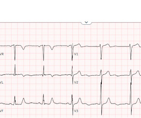
This certainly looks like an anterior STEMI (proximal LAD occlusion), with STE and hyperacute T-waves (HATW) in V2-V6 and I and aVL. How do you explain the anterior STEMI(+)OMI immediately after ROSC evolving into posterior OMI 30 minutes later? This caused a type 2 anterior STEMI.
This was a machine read STEMI positive OMI. The meaning of this quote is that at times, something as obvious as the dramatic anterior lead ST elevation that we see in today's tracing is not the result of an acute LAD STEMI. His ECG is shown below. Pretty obvious anterior current of injury. What would you guess is the culprit artery?
See this post: Septal STEMI with ST elevation in V1 and V4R, and reciprocal ST depression in V5, V6. After being transferred to an academic center, she was taken to the cath lab: Proximal RCA occlusion (causing inferior and RV OMI) Unfortunately, she continued to decline despite aggressive measures. Also seen in inferior + RV OMI.)
In the available view of the sinus rhythm, we see normal variant STE which probably meets STEMI criteria in V4 and V5. In other words, the inferior "ST elevation" is due to the abnormal rhythm, and does not signify OMI or STEMI in any way. This situation has been named "Emery phenomenon." YOU TOO CAN HAVE THE PM Cardio AI BOT!!
Recall from this post referencing this study that "reciprocal STD in aVL is highly sensitive for inferior OMI (far better than STEMI criteria) and excludes pericarditis, but is not specific for OMI." Immediate versus delayed invasive intervention for non-stemi patients. Academic Emergency Medicine 27(S1): S220; May 2020.
He called 911 and paramedics recorded a prehospital 12 lead ECG which showed a clear inferior STEMI (not shown, tracing could not be found). Published in Academic Emergency Medicine, vol. Objectives : To find the incidence of any rSTD or T-wave inversion (TWI) in angiographically proven inferior STEMI.
Post Cath ECG: Obviously completing MI with LVA morphology, and STE that meets STEMI criteria (but pt is still diagnosed as "NSTEMI"). Day 12 ECG: FINAL DIAGNOSIS: "NSTEMI" Despite the fact that his day 4 ECG easily meets STEMI criteria, the patient is diagnosed as NSTEMI. Academic Emergency Medicine 27(S1): S220; May 2020.
There is an obvious inferior posterior STEMI(+) OMI. We recorded an ECG in which V1-V3 were put in the position of V4R-V6R, and V4-6 were placed in V7-9 to (academically) confirm posterior OMI. I say academically because the STD in V2 is diagnostic -- posterior leads are NOT necessary. What is the atrial activity? What to do?
We found that 38% of out of hospital ventricular fibrillation was due to STEMI. Correlation of STEMI in Resuscitated Non-traumatic out-of-hospital Cardiopulmonary Arrest patients with Initial Rhythm and Cardiac Catheterization Findings (Abstract 580). Academic Emergency Medicine 17(s1):S194; May 2010 Reference : Scott NL.
It is a sad academic story ,most of the interventional cardiology community shrugged it off as a non-event. No one knows how the pPCI related delay was legally ratified and academically accepted by the elite cardiology forums. In LAD STEMI time is more crucial. In many countries, the hub-and-spoke model is struggling to deliver.
The primary safety end point was Bleeding Academic Research Consortium 3 to 5 bleeding at 30 days.RESULTS:Between January 10, 2019, and September 18, 2021, a total of 2989 patients were randomized. The primary efficacy end point occurred in 37 patients (2.5%) in both the PPA and placebo groups (hazard ratio, 1.00 [95% CI, 0.63
This is diagnostic of inferior MI, though does not meet millimeter criteria for "STEMI." He was worried for inferior MI and ordered another, which was recorded 15 minutes later: Now clearly and obviously diagnostic of inferior STEMI. Academic Emergency Medicine 24(1):120-124. It is not subtle any more. References : 1.
This was marked as "Not a STEMI" by the physicians. It is not a STEMI, but it is diagnostic of an LAD OMI (Occlusion MI). has outperformed many cardiologists in its ability to recognize with "high confidence" acute OMIs from ECGs not satisfying STEMI-criteria. This was sent to me by a friend.
The ECG shows obvious STEMI(+) OMI due to probable proximal LAD occlusion. Distinction of PMVT vs VFib is an academic one in this case ). The patient in today’s case is a previously healthy 40-something male who contacted EMS due to acute onset crushing chest pain. The below ECG was recorded. Both PMVT and VFib occurred multiple times.
They argue for either immediate intervention or defer transiently, postpone or just ignore , based on clinical ,hemodynamic * , Individual, institutional , or some other non academic factors. After all, globally 90% of all successful myocardial reperfusion is done by the humble streptokinase or the more glamorous TNK -TPA.
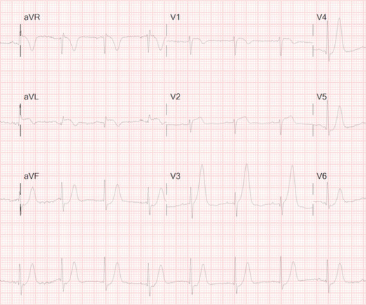
It doesn’t meet any conventional STEMI criteria, but there is patently obvious increased area under the curve. 2] Exotic ECG findings – in this case, PR-interval shortening – make for excellent academic inquiry, but should never be a point of distraction from pathognomonic occlusive coronary disease. Is this OMI?
Still , even today it is weird to see hours of academic time is consumed in CTO Interventions in any interventional cardiology meets. One more remote risk in CTO is, acute collateral shutdown causing STEMI/NSTEMI. Surprise… surprise !, There is some good news. What will happen if a single donor (RCA/LCX) gets closed?
The axiom of "type 1 (ACS, plaque rupture) STEMIs are not tachycardic unless they are in cardiogenic shock" is not applicable outside of sinus rhythm. Is that an obvious STEMI underneath that rhythm? Is this inferor STEMI? Atrial Flutter with Inferior STEMI? If I fix the rhythm will the ST changes resolve?
Still does not meet STEMI criteria, but it is an obvious OMI And then another one became more obvious: Cath lab was activated and a 100% RCA occlusion was found. There is no ST elevation in inferior leads, but it may just be too early to manifest. Learning Points: 1. Beware ST depression in I and aVL.
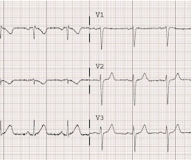
There is mixed overlap of ST-segment elevation (STE), ST-segment depression (STD), Hyperacute T waves (HATW), and deWinter pattern (which the ACC regards as a STEMI-equivalent but is better suited under the blanket of OMI). mg of SL NTG, along with a total of 100 mcg of Fentanyl with little to no change in symptoms.
The ECG was read as "No STEMI" and the patient was treated like an average chest pain patient (despite the fact that a chest pain patient with active pain and active subendocardial ischemia is very high risk). Academic Emergency Medicine 27(S1): S220; May 2020.
Code STEMI was activated. A man in his 80s with chest pain What, besides large anterior STEMI, is so ominous about this ECG? From an academic standpoint — I found several subtle ECG findings from the 2 tracings in this case to be especially interesting. This one of the highest risk OMI patterns possible to see on an ECG.
Immediate and early percutaneous coronary intervention in very high-risk and high-risk Non-STEMI patients. Academic Emergency Medicine 27(S1): S220. Unfortunately, they follow their own guidelines only 6% of the time!! Lupu L, et al. Clin Cardiol 2022; [link] Labs included: hsTnI 156 ng/L, Hb 12 g/dL, WBC 12x10^9/L, Cr. mg/dL, K 3.5
He has a history of coronary artery disease and a STEMI two years prior that was treated with primary PCI. At the time of this initial ED ECG, his symptoms were improving ECG #1 on admission to the ED The patient was not seen quickly in the ED as it was a busy shift and the ECG did not meet STEMI criteria. The below ECG was recorded.
It is a strange academic habit among cardiologists, that they have subdivided medical management into optimal and suboptimal. He is a STEMI patient (1 year old) with mild LV dysfunction and thinning of IVS and anterior wall. Academic lessons from this patient.
We organize all of the trending information in your field so you don't have to. Join thousands of users and stay up to date on the latest articles your peers are reading.
You know about us, now we want to get to know you!
Let's personalize your content
Let's get even more personalized
We recognize your account from another site in our network, please click 'Send Email' below to continue with verifying your account and setting a password.


Let's personalize your content