This site uses cookies to improve your experience. To help us insure we adhere to various privacy regulations, please select your country/region of residence. If you do not select a country, we will assume you are from the United States. Select your Cookie Settings or view our Privacy Policy and Terms of Use.
Cookie Settings
Cookies and similar technologies are used on this website for proper function of the website, for tracking performance analytics and for marketing purposes. We and some of our third-party providers may use cookie data for various purposes. Please review the cookie settings below and choose your preference.
Used for the proper function of the website
Used for monitoring website traffic and interactions
Cookie Settings
Cookies and similar technologies are used on this website for proper function of the website, for tracking performance analytics and for marketing purposes. We and some of our third-party providers may use cookie data for various purposes. Please review the cookie settings below and choose your preference.
Strictly Necessary: Used for the proper function of the website
Performance/Analytics: Used for monitoring website traffic and interactions
Cath lab declined as it is not a STEMI." And now this finding is even formally endorsed as a "STEMI equivalent" in the 2022 ACC guidelines!!! Another myocardial wall is sacrificed at the altar of the STEMI/NonSTEMI mindset. Opiates are associated with worse outcomes in MyocardialInfarction. Abstract 556.
Triage ECG (no prior for comparison): Computer algorithm read: "Sinus rhythm, low voltage QRS, inferior myocardialinfarction, probably old." Post Cath ECG: Obviously completing MI with LVA morphology, and STE that meets STEMI criteria (but pt is still diagnosed as "NSTEMI"). Academic Emergency Medicine 27(S1): S220; May 2020.
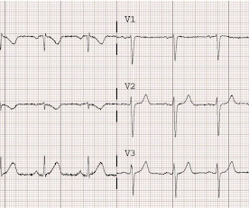
There is an obvious inferior posterior STEMI(+) OMI. We recorded an ECG in which V1-V3 were put in the position of V4R-V6R, and V4-6 were placed in V7-9 to (academically) confirm posterior OMI. I say academically because the STD in V2 is diagnostic -- posterior leads are NOT necessary. What is the atrial activity? What to do?
CAPTIM trial was published in 2002, which left a gospel truth in the science of myocardial reperfusion (two decades gone now). It is a sad academic story ,most of the interventional cardiology community shrugged it off as a non-event. In LAD STEMI time is more crucial. This is what CAPTIM told us. NRMI data Ref 2) 2.There
He called 911 and paramedics recorded a prehospital 12 lead ECG which showed a clear inferior STEMI (not shown, tracing could not be found). Published in Academic Emergency Medicine, vol. Objectives : To find the incidence of any rSTD or T-wave inversion (TWI) in angiographically proven inferior STEMI.
This is diagnostic of inferior MI, though does not meet millimeter criteria for "STEMI." He was worried for inferior MI and ordered another, which was recorded 15 minutes later: Now clearly and obviously diagnostic of inferior STEMI. Academic Emergency Medicine 24(1):120-124. It is not subtle any more. References : 1.
He denied any known medical history, specifically: coronary artery disease, hypertension, dyslipidemia, diabetes, heart failure, myocardialinfarction, or any prior PCI/stent. It doesn’t meet any conventional STEMI criteria, but there is patently obvious increased area under the curve. No appreciable skin pallor. Is this OMI?
The ECG was read as "No STEMI" and the patient was treated like an average chest pain patient (despite the fact that a chest pain patient with active pain and active subendocardial ischemia is very high risk). A New ST-segment elevation myocardialinfarction equivalent pattern? Eur J Emerg Med. 2017;24:236–242. Am J Emerg Med.
Occlusion myocardialinfarction is a clinical diagnosis Written by Willy Frick (@Willyhfrick). Recall from this post referencing this study that "reciprocal STD in aVL is highly sensitive for inferior OMI (far better than STEMI criteria) and excludes pericarditis, but is not specific for OMI." The case continues. Worrall, C.,
Immediate and early percutaneous coronary intervention in very high-risk and high-risk Non-STEMI patients. Association between opioid analgesia and delays to cardiac catheterization of patients with occlusion MyocardialInfarctions. Academic Emergency Medicine 27(S1): S220. Lupu L, et al. mg/dL, K 3.5 Abstract 556.
Patients with ST-segment–elevation myocardialinfarction undergoing primary percutaneous coronary intervention were randomly assigned by center to receive low-dose PPA or matching placebo for at least 48 hours. mg·kg·h of bivalirudin intravenously). mg·kg·h of bivalirudin intravenously).
See this post: Septal STEMI with ST elevation in V1 and V4R, and reciprocal ST depression in V5, V6. After being transferred to an academic center, she was taken to the cath lab: Proximal RCA occlusion (causing inferior and RV OMI) Unfortunately, she continued to decline despite aggressive measures. Also seen in inferior + RV OMI.)
The ECG shows obvious STEMI(+) OMI due to probable proximal LAD occlusion. Troponin T peaked at 38,398 ng/L ( = a very large myocardialinfarction, but not massive-- thanks to the pre-PCI spontaneous reperfusion, and rapid internvention!! ). Distinction of PMVT vs VFib is an academic one in this case ).
The axiom of "type 1 (ACS, plaque rupture) STEMIs are not tachycardic unless they are in cardiogenic shock" is not applicable outside of sinus rhythm. Is that an obvious STEMI underneath that rhythm? Is this inferor STEMI? Atrial Flutter with Inferior STEMI? If I fix the rhythm will the ST changes resolve?
Code STEMI was activated. A man in his 80s with chest pain What, besides large anterior STEMI, is so ominous about this ECG? Primary angioplasty in acute myocardialinfarction with right bundle branch block: should new onset right bundle branch block be added to future guidelines as an indication for reperfusion therapy?
He has a history of coronary artery disease and a STEMI two years prior that was treated with primary PCI. At the time of this initial ED ECG, his symptoms were improving ECG #1 on admission to the ED The patient was not seen quickly in the ED as it was a busy shift and the ECG did not meet STEMI criteria. The below ECG was recorded.
The paramedics diagnosis was "Possible Anterolateral STEMI." More proof that a huge STEMI may have normal or near normal initial troponin. Taken together, these findings suggest an ongoing extensive antero-lateral STEMI. I don't know what the device algorithm interpretation stated. The final angiographic result is very good.
We organize all of the trending information in your field so you don't have to. Join thousands of users and stay up to date on the latest articles your peers are reading.
You know about us, now we want to get to know you!
Let's personalize your content
Let's get even more personalized
We recognize your account from another site in our network, please click 'Send Email' below to continue with verifying your account and setting a password.


Let's personalize your content