This site uses cookies to improve your experience. To help us insure we adhere to various privacy regulations, please select your country/region of residence. If you do not select a country, we will assume you are from the United States. Select your Cookie Settings or view our Privacy Policy and Terms of Use.
Cookie Settings
Cookies and similar technologies are used on this website for proper function of the website, for tracking performance analytics and for marketing purposes. We and some of our third-party providers may use cookie data for various purposes. Please review the cookie settings below and choose your preference.
Used for the proper function of the website
Used for monitoring website traffic and interactions
Cookie Settings
Cookies and similar technologies are used on this website for proper function of the website, for tracking performance analytics and for marketing purposes. We and some of our third-party providers may use cookie data for various purposes. Please review the cookie settings below and choose your preference.
Strictly Necessary: Used for the proper function of the website
Performance/Analytics: Used for monitoring website traffic and interactions
tim.hodson Wed, 04/16/2025 - 14:19 April 16, 2025 An artificial intelligence (AI) program trained to review images from a common medical test can detect early signs of tricuspid heart valve disease and may help doctors diagnose and treat patients sooner, according to research from the Smidt Heart Institute at Cedars-Sinai.
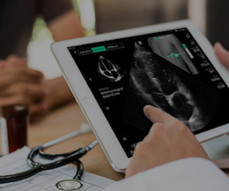
tim.hodson Tue, 04/15/2025 - 15:54 April 15, 2025 DESKirecently announced FDA clearance for HeartFocus , its AI-enabled heart exam software that empowers any healthcare professional, including novices, to perform clinical-quality heart scans from any compatible device after just a few hours of training.
tim.hodson Tue, 02/18/2025 - 16:17 Feb. 11, 2025 UltraSight, a company committed to enhancing the efficiency and productivity of cardiac ultrasound,recentlyannounced support from Bristol Myers Squibb (BMS) for a study that aims to improve access to echocardiographic assessments for patients with obstructive hypertrophic cardiomyopathy (oHCM).
Sectra Cardiology Imaging addresses the long-standing challenge of fragmented cardiac imaging by unifying ECGs, Echo, Cath, Cardiac NM, Vascular, and CT FFR within a single, intuitive interface.
Stroke, Volume 56, Issue Suppl_1 , Page ATMP19-ATMP19, February 1, 2025. If indicated, the patient will be scheduled for an MRI within 24 hours and/or an echocardiogram within 48 hours of the TIA clinic visit. Radiology and cardiology have availability for urgent MRIs and echocardiograms.
Stroke, Volume 56, Issue Suppl_1 , Page AWP44-AWP44, February 1, 2025. Residual shunt post PFO closure was assessed using transthoracic echocardiogram (TTE) with saline contrast. Background:Patent foramen ovale (PFO) is an independent risk factor for neurovascular injury such as stroke.
Stroke, Volume 56, Issue Suppl_1 , Page ATP182-ATP182, February 1, 2025. Advanced cardiac workup (ACW), including transesophageal echocardiogram (TEE) and implantable loop recorder (ILR) are widely considered a crucial element in the ESUS work-up. The etiology of AChA infarcts remains poorly understood.
Circulation: Cardiovascular Quality and Outcomes, Volume 18, Issue 1 , Page e011504, January 1, 2025. BACKGROUND:Risk stratification strategies for cancer therapeuticsrelated cardiac dysfunction (CTRCD) rely on serial monitoring by specialized imaging, limiting their scalability. A high-risk versus low-risk AI-ECG screen (0.1 fold and 13.5-fold
tim.hodson Mon, 03/03/2025 - 12:09 March 3, 2025 Eko Health has published a peer-reviewed study in JACC Advances evaluating its FDA-cleared AI model for detecting reduced ejection fraction (EF), a common marker of a weakened heart that can't pump blood effectively (also known as heart failure with reduced ejection fraction, HFrEF).
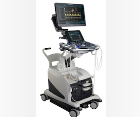
tim.hodson Mon, 03/24/2025 - 11:46 March 24, 2025 FUJIFILM Healthcare Americas Corp.and Us2.ai ais software, when used with the LISENDO 880 ultrasound system, fully automates the analysis and reporting of echocardiograms and provides comprehensive cardiac measurements for the diagnosis of heart disease.
Despite apparently hearing the above history together with two diagnostic ECGs and a troponin compatible with OMI, the cardiologist thought the ECG represented pericarditis and recommended echocardiogram. Echocardiogram was finally performed five hours after the first diagnostic ECG. This far out, the benefit of PCI is very attenuated.
Long term outcome is not available. == MY Comment, by K EN G RAUER, MD ( 2/1/2025 ): == We need to learn from cases like today's. Total proximal LAD occlusion was found and stented at angiography soon after the ECG above. Here are some images: Next morning ECG: Reperfusion findings are clear.
Echocardiogram showed inferior hypokinesis. As you can see, the lesion is not very angiographically impressive , more on this below. Nevertheless, the operator performed intravascular ultrasound and saw erupted calcium nodule consistent with plaque erosion. Troponin was rising when last checked, 8928 ng/L.
More troponin values were measured at the cardiac center: 2327- 267 ng/L 0821- 355 ng/L 1108- 305 ng/L An echocardiogram on day three of the patients admission showed an ejection fraction of 46% with abnormal basal inferior and basal lateral segments, and severe aortic stenosis.
In this study of consecutive patients with LBBB who were hospitalized and had an echocardiogram, a QRS duration less than 170 ms (n = 262), vs. greater than 170 ms (n = 38), was associated with a significantly better ejection fraction (36% vs. 24%). So indeed the QRS is approximately 200 ms. Comment: What is the normal QRS duration in LBBB?
It is reasonable to perform an echocardiogram to evaluate LV function. Catheter ablation or flecainide should be considered in symptomatic patients with idiopathic VT/PVCs from an origin other than the RVOT or the left fascicles. [ 1 ] Considerations Regarding Use of Flecainide: A 12-lead ECG is mandatory before starting therapy.
An echocardiogram at 13:40 showed: Severely reduced global systolic function with an estimated EF of 10-20% Mildly increased LV size Akinesis of the entire septum and apex Hypokinesis of the anterior, anterolateral, and mid posterior segments A final troponin T was drawn at 17:23- 3,475 ng/L.
We organize all of the trending information in your field so you don't have to. Join thousands of users and stay up to date on the latest articles your peers are reading.
You know about us, now we want to get to know you!
Let's personalize your content
Let's get even more personalized
We recognize your account from another site in our network, please click 'Send Email' below to continue with verifying your account and setting a password.


Let's personalize your content