This site uses cookies to improve your experience. To help us insure we adhere to various privacy regulations, please select your country/region of residence. If you do not select a country, we will assume you are from the United States. Select your Cookie Settings or view our Privacy Policy and Terms of Use.
Cookie Settings
Cookies and similar technologies are used on this website for proper function of the website, for tracking performance analytics and for marketing purposes. We and some of our third-party providers may use cookie data for various purposes. Please review the cookie settings below and choose your preference.
Used for the proper function of the website
Used for monitoring website traffic and interactions
Cookie Settings
Cookies and similar technologies are used on this website for proper function of the website, for tracking performance analytics and for marketing purposes. We and some of our third-party providers may use cookie data for various purposes. Please review the cookie settings below and choose your preference.
Strictly Necessary: Used for the proper function of the website
Performance/Analytics: Used for monitoring website traffic and interactions
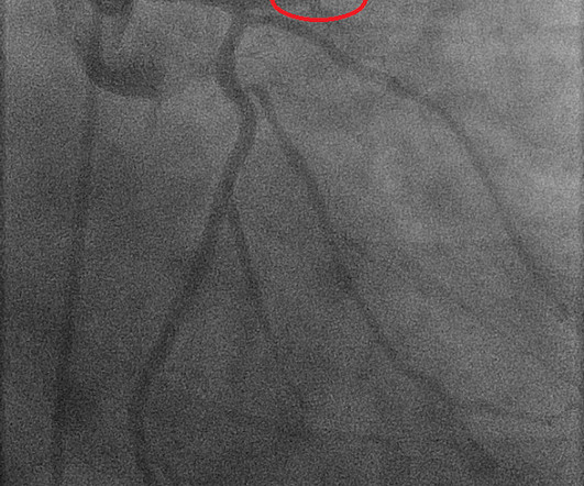
Coronary artery spasm (CAS), or Prinzmetal angina, is a recognised cause of myocardial ischaemia in non-obstructed coronary arteries which typically presents with anginal chestpain. The patient presented with recurrent palpitations and pre-syncope, with no chestpain.
Written by Hans Helseth A 34 year old man with no known medical history presented to the ED after an hour of chestpain. He described the pain as a mid sternal "burning sensation" and rated it 8.5 out of 10 at onset, but on presentation to the ED, reported that the pain had improved to 4.5. 10 chestpain.
Ventricular tachycardia?) The patient received three nitroglycerin tablets with significant "improvement" in his chestpain. Improved chestpain is unresolved chestpain. I am commonly told, and I commonly read in the chart that chestpain is resolved. What do you think? Proximal LAD."
A 69 year old woman with a history of hypertension presented to the emergency department by EMS for evaluation of chestpain and shortness of breath. She awoke in the morning with sharp chestpain which worsened throughout the morning. As her pain worsened, so did her dyspnea. This was written by Hans Helseth.
See these other related cases: Acute chestpain and ST Elevation. A woman in her 20s with syncope 80-something year old with acute chestpain. CT done to look for aortic dissection. Although other entities may produce various forms of alternans ( as discussed in the Oct.
He denied chestpain. A Chest X-ray did not show pulmonary edema. This ECG was recorded: It is difficult to appreciate P-waves, but I believe this is sinus tachycardia. It is correct that he did not have chestpain, but we must remember that fully 1/3 of full blown STEMI do not present with chestpain.
In view of the clinical history ( ie, that this patient had a history of longstanding AFlutter but as far as we know, no chestpain ) this ST-T wave change most likely reflects a "memory" effect , in which there will often be ST-T wave abnormalities that persist for hours ( up to a few days ) following a long period of a sustained tachycardia.
The ECG in Figure-1 was obtained from a middle-aged man who presents to the ED ( E mergency D epartment ) with 6 hours of chestpain. Figure-1: The initial ECG in today's case obtained from a middle-aged man with 6 hours of chestpain. ( He is hemodynamically stable. However, Atropine is not benign.
A middle-age woman with no previous cardiac history called 911 for chestpain. This was her prehospital ECG: What do you think? There is sinus rhythm with RBBB and obvious LAD OMI (proximal LAD occlusion): hyperacute T-waves in I, aVL and minimal STE in V1, V2. DOI: 10.1016/j.resuscitation.2025.110515
A 50-something male presented to triage with chestpain for one day. A Chest X-ray showed infiltrates. Thus, another etiology of chestpain is found, and the fever suggests "fever-induced Brugada." The presenting complaint noted at Triage was, "a 50yo man with chestpain!" The temperature was 39.5
He played a round of golf a week prior and felt an episode of chestpain during the round, which spontaneously resolved. On presentation, he reported no chestpain or shortness of breath. Five days later, the patient was exercising when he developed chestpain at 19:30 which lasted for an hour. Or both?
Written by Pendell Meyers An adult man presented with acute chestpain. It is a wide complex regular tachycardia at a rate of 120. Is it ventricular tachycardia? It is ofen downsloping This one is also a wide complex tachycardia. He appeared critically ill. Near 100% mortality without rapid reperfusion."
Sent by anonymous, written by Pendell Meyers A woman in her 40s with no known comorbidities presented with acute chestpain radiating to left arm and neck, which started approximately 4 hours prior to arrival. Vitals were reported as within normal limits except for tachycardia. 68 minutes with chest compressions, full recovery.
Smith comments : Wide complex tachycardia. The differential diagnosis of WCT is: 1) Sinus tachycardia with "aberrancy" (in this case RBBB and LAFB), but there are no P-waves and the QRS morphology is not typical of simple RBBB/LAFB. Also, if the rate is constant, not wavering up and down, it is highly unlikely to be sinus tachycardia.
He had no chestpain, dyspnea, or any other anginal equivalent, and his vital signs were normal. This was overtaken by a predominance of sympathetic surge ( tachycardia, persistent ST elevation development of electrical "storm" with failure to respond to recurrent defibrillation ). & Dawson, D.
We organize all of the trending information in your field so you don't have to. Join thousands of users and stay up to date on the latest articles your peers are reading.
You know about us, now we want to get to know you!
Let's personalize your content
Let's get even more personalized
We recognize your account from another site in our network, please click 'Send Email' below to continue with verifying your account and setting a password.


Let's personalize your content