This site uses cookies to improve your experience. To help us insure we adhere to various privacy regulations, please select your country/region of residence. If you do not select a country, we will assume you are from the United States. Select your Cookie Settings or view our Privacy Policy and Terms of Use.
Cookie Settings
Cookies and similar technologies are used on this website for proper function of the website, for tracking performance analytics and for marketing purposes. We and some of our third-party providers may use cookie data for various purposes. Please review the cookie settings below and choose your preference.
Used for the proper function of the website
Used for monitoring website traffic and interactions
Cookie Settings
Cookies and similar technologies are used on this website for proper function of the website, for tracking performance analytics and for marketing purposes. We and some of our third-party providers may use cookie data for various purposes. Please review the cookie settings below and choose your preference.
Strictly Necessary: Used for the proper function of the website
Performance/Analytics: Used for monitoring website traffic and interactions
Methods We systematically searched PubMed/MEDLINE, EMBASE and Cochrane CENTRAL from inception to 22 January 2024. They included randomised controlled trials that enrolled adults undergoing major cardiac surgeries and reported postpericardiotomy syndrome, pericardial effusion and pericarditis as primary or secondary outcomes.
Overall, this looks like one of the rare ECGs that is actually specific for pericarditis in my opinion. Pericarditis maybe." These include the following: i ) Today's case provides an example of an initial ECG, that in Dr. Meyers' words — "is one of the rare ECGs that is actually specific for pericarditis".
DALLAS, June 17, 2024 — About 40,000 people in the United States experience recurrent pericarditis, or inflammation of the sac-like structure that protects the heart, which can cause chest pain and may lead to fluid buildup around the heart muscle.
The computer interpretation was “ST elevation, consider early repolarization, pericarditis or injury.” The final cardiology interpretation confirmed the computer interpretation of “ST elevation, consider early repolarization, pericarditis or injury”. A healthy 45-year-old female presented with chest pain, with normal vitals.
Acute pericarditis is characterized by pericardial inflammation which can be treated with anti-inflammatory drugs. A considerable percentage of patients develops recurrent pericarditis with several relapses. Two pathophysiological mechanisms have been described for idiopathic recurrent pericarditis, autoimmune and autoinflammatory.
Publication date: Available online 20 May 2024 Source: The American Journal of Cardiology Author(s): Shaye Kivity, Tomer Ziv Baran, Miri Mizrahi Reuveni, Angela Irony, Limor Adler, Yehuda Alder, Roma Parikh, Sara Kivity
This is a value typical for a large subacute MI, n ormal value 48 hours after myocardial infarction is associated with Post-Infarction Regional Pericarditis ( PIRP ). As already mentioned, this patient could have post-infarction regional pericarditis from a large completed MI. Sinus tachycardia has many potential causes. Hammill SC.
The undergraduate continues: This new EKG pattern is more suggestive of acute pericarditis. Usually with pericarditis, some degree of PR segment depression is expected. This is typical of pericarditis. But, as I always say, you diagnose pericarditis at your peril. This EKG seems to lack it.
Publication date: Available online 10 December 2024 Source: The American Journal of Cardiology Author(s): Rehan Karmali, Issam Motairek, Samia Mazumder, Felix Berglund, Lorenzo Braghieri, Astefanos Al-Dalakta, Katherine Singh, Brittany Weber, Allan Klein
It is easy to say pericarditis in such a case. young male no risk factors and ST-elevation in several leads) As Dr. Smith has emphasized many times you diagnose pericarditis at your patient's and your own peril. Version 1 was not trained to detect myo- or pericarditis. The above ECG was recorded. How did the Queen do?
milla1cf Thu, 03/28/2024 - 07:28 March 28, 2024 — Biosense Webster, Inc., patients, were presented as a late-breaker at the 2024 AF Symposium. In Europe, the TRUPULSE generator received CE mark in late 2023 and the VARIPULSE Catheter received CE mark in February 2024. Food & Drug Administration ( FDA ). In the U.S.,
Int J Cardiol 2024 2. ST depression in lead AVL differentiates inferior ST-elevation myocardial infarction from pericarditis. Eur Herat J Digital Health 2024 OMI ECG findings can lead to rapid diagnosis, and can be widely disseminated through AI References 1. De Alencar Neto. Nikus et al. Curr Cardiol Red 2021 3. Kontos et al.
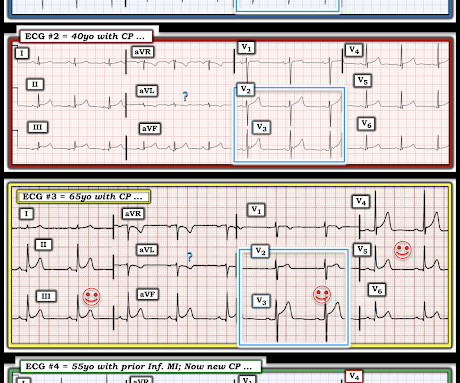
These latter findings are typical of pericarditis, but pericarditis never has reciprocal ST depression. Usually with pericarditis and myocarditis — hyperacute T waves (HATW) are not present. S mith : there is STE in lead III and reciprocal STD in aVL. This is OMI until proven otherwise.
Accessed May 28, 2024. Dyspnea, Chest pain, Tachypneic, Ill appearing: Bedside Cardiac Echo gives the Diagnosis 31 Year Old Male with RUQ Pain and a History of Pericarditis. Electrocardiographic Differentiation Between Acute Pulmonary Embolism and Acute Coronary Syndromes on the Basis of Negative T Waves - ScienceDirect.
Circulation, Volume 150, Issue Suppl_1 , Page A4139677-A4139677, November 12, 2024. Postoperative recovery was successful, with no further hemoptysis, and the patient was discharged to a rehabilitation facility.Discussion:EPDs are no longer used due to high failure rates and complications like scar formation and constrictive pericarditis.
ECG of pneumopericardium and probable myocardial contusion shows typical pericarditis Male in 30's, 2 days after Motor Vehicle Collsion, complains of Chest Pain and Dyspnea Head On Motor Vehicle Collision. : A Child with Blunt Trauma -- See how the ECG can be definite for myocardial contusion, but subtle, and what happens if you miss it.
Then the patient's pain then resolved spontaneously after 2 sublingual nitroglycerine and another ECG was recorded ECG 2 at 16 minutes ST ELEVATION CONSISTENT WITH INJURY, PERICARDITIS, OR EARLY REPOLARIZATION Overread same Smith : The T-waves are now MUCH smaller. The S-wave is reconstituted. The inferior findings are much less pronounced.
You can easily imagine this patient getting one of several diagnoses -- vasospasm, MINOCA , pericarditis, or maybe even no diagnosis at all beyond "non-obstructive coronary artery disease." In a large proportion of cath labs, the operator would probably have ended the case at this point.
The "flu-like" illness suggests myo- or pericarditis, but that would be a diagnosis of exclusion. The scale is wrong. == MY Comment , by K EN G RAUER, MD ( 1/14/ 2024 ): == I was not taught about artifact in either medical school or during my residency. Do not wait for the troponin; a lot of myocardium will be dead if you do.
Circulation, Volume 150, Issue Suppl_1 , Page A4141403-A4141403, November 12, 2024. To-date no larger studies have assessed sub-clinical myocardial mechanics in acute tuberculous pericarditis (ATBP) and age/sex/ethnic differences, and synergistic-prognostic association of these parameters with patients' outcomes (survival).Aim:To
The initial computer and final cardiology interpretation was a differential: “ST elevation, consider early repolarization, pericarditis, or injury.” But STEMI criteria ignore all this and look at ST segments in isolation. Based on STEMI criteria and unhelpful computer interpretation, the patient was rushed to the cath lab.
The exception is with postinfarction pericarditis , in which a completed transmural infarct results in inflammation of the subepicardial myocardium and STE in the distribution of the infarct, and which results in increased STE and large upright T-waves. These findings together are more commonly seen with pericarditis.
In addition, ischemic STD in aVL is highly sensitive for inferior OMI, and excludes pericarditis. for both pericarditis and normal variant, the vector results in more STE in II than III, and absence of STD in aVL. It means that, if there is STD in aVL, then it is almost certainly not pericarditis (but could be myocarditis).
Dyspnea, Chest pain, Tachypneic, Ill appearing: Bedside Cardiac Echo gives the Diagnosis 31 Year Old Male with RUQ Pain and a History of Pericarditis. Submitted by a Med Student, with Great Commentary on Bias! Chest pain, SOB, Precordial T-wave inversions, and positive troponin. What is the Diagnosis?
See here for young women with OMI == MY Comment , by K EN G RAUER, MD ( 12/24 /2024 ): == I found today's case challenging and an excellent example of how despite my not being certain of the diagnosis from the history and initial ECG careful follow-up yielded the answer. Smith : I recognize this as a STEMI mimic. I was not alarmed.
We organize all of the trending information in your field so you don't have to. Join thousands of users and stay up to date on the latest articles your peers are reading.
You know about us, now we want to get to know you!
Let's personalize your content
Let's get even more personalized
We recognize your account from another site in our network, please click 'Send Email' below to continue with verifying your account and setting a password.


Let's personalize your content