This site uses cookies to improve your experience. To help us insure we adhere to various privacy regulations, please select your country/region of residence. If you do not select a country, we will assume you are from the United States. Select your Cookie Settings or view our Privacy Policy and Terms of Use.
Cookie Settings
Cookies and similar technologies are used on this website for proper function of the website, for tracking performance analytics and for marketing purposes. We and some of our third-party providers may use cookie data for various purposes. Please review the cookie settings below and choose your preference.
Used for the proper function of the website
Used for monitoring website traffic and interactions
Cookie Settings
Cookies and similar technologies are used on this website for proper function of the website, for tracking performance analytics and for marketing purposes. We and some of our third-party providers may use cookie data for various purposes. Please review the cookie settings below and choose your preference.
Strictly Necessary: Used for the proper function of the website
Performance/Analytics: Used for monitoring website traffic and interactions
Written by Jesse McLaren A 45-year-old presented with 24 hours of intermittent chestpain. On it’s own this is nonspecific, but in the right context this could be diagonal occlusion (if active chestpain) or infero-posterior reperfusion (if resolved chestpain). #2 Can you guess the sequence?
I assumed it was a patient with acute chestpain. It was a man in his 30s with chestpain. Click here to sign up for Queen of Hearts Access == MY Comment , by K EN G RAUER, MD ( 10/1 /2024 ): == I looked at the ECG in today’s case knowing only that the patient was a younger male adult with CP ( C hest P ain ).
Written by Jesse McLaren, with a very few edits by Smith A 60-year-old presented with chestpain. Inferior hyperacute T waves, which have been added to the 2022 ACC consensus on chestpain as a “STEMI equivalent”[3] 3. Int J Cardiol 2024 2. Eur Herat J Digital Health 2024 Nikus et al. Kontos et al.
Written by Colin Jenkins and Nhu-Nguyen Le with edits by Willy Frick and by Smith A 46-year-old male presented to the emergency department with 2 days of heavy substernal chestpain and nausea. The patient continued having chestpain. These diagnoses were not found in his medical records nor even a baseline ECG.
Written by Pendell Meyers A man in his 40s called EMS for acute chestpain that awoke him from sleep, along with nausea and shortness of breath. DO NOT MISS THESE! == MY Comment , by K EN G RAUER, MD ( 9/7 /2024 ): == The KEY consideration in today's case is — How effectively can we diagnose acute OMI in a patient with RBBB?
Written by Pendell Meyers A man in his 60s presented with acute chestpain and normal vital signs. Here is his triage ECG: What do you think? The ECG shows massively hyperacute T waves of LAD OMI, plus WPW. V3-V5 also have the depressed HATW takeoff which qualifies them as the rare de Winter subtype of HATWs. 13 post.
Written by Jesse McLaren A healthy 75 year old developed 7/10 chestpain associated with diaphoresis and nausea, which began on exertion but persisted. Below is the first ECG recorded by paramedics after 2 hours of chestpain, interpreted by the machine as “possible inferior ischemia”. What do you think?
By Magnus Nossen This ECG is from a young man with no risk factors for CAD, he presented with chestpain. The patient is a young adult male with chestpain. The chestpain was described as pressure like and radiation to both arms and the jaw. (THE How would you assess this ECG? What is your next step?
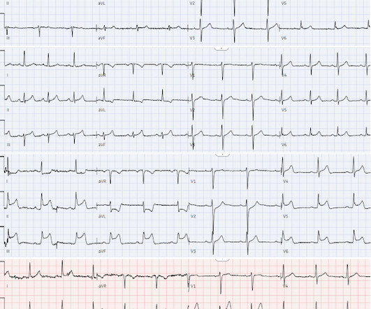
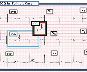
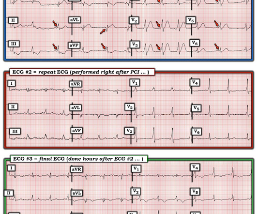
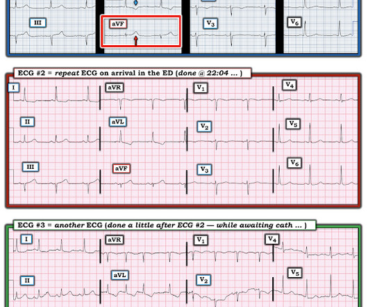
Written by Magnus Nossen with Edits by Grauer and Smith The ECGs in today’s case are from 3 different patients all presenting with new-onset CP ( ChestPain ). All ECGs were recorded by EMS, and transferred to a PCI capable center for evaluation. For 2 of the 3 patients — the cath lab was activated based on the ECG.
No ChestPain, but somnolent. The fact that this is syncope makes give it a far lower pretest probability than chestpain, but it was really more than syncope, as the patient actually underwent CPR and had hypotension on arrival of EMS. Here is the ED ECG (a photo of the paper printout) What do you think?
A 50-something male with hypertension and 20- to 40-year smoking history presented with 1 week of stuttering chestpain that is worse with exertion, which takes many minutes to resolve after resting and never occurs at rest. At times the pain does go to his left neck. It is a ssociated with mild dyspnea on exertion.
I was working at triage when the medics brought this patient who is 65 yo and has had chestpain for 12 hours. Click here to sign up for Queen of Hearts Access == MY Comment , by K EN G RAUER, MD ( 7/20 /2024 ): == Today's case illustrates the importance of attention to subtle serial ECG findings.
Written by Willy Frick A man in his 50s with history of hypertension, hyperlipidemia, and a 30 pack-year smoking history presented to the ER with 1 hour of acute onset, severe chestpain and diaphoresis. His ECG is shown: What do you think? What do you think?
Written by Pendell Meyers A man in his late 30s with history of hypertension, tobacco use, and obesity presented to the Emergency Department for acute chestpain which started approximately 3 hours prior to arrival, in the setting of a very stressful situation. The pain radiated down both arms, 10/10 in severity.
He did not remember whether he had experienced any chestpain. Within a few days, the patient was extubated and was neurologically intact. However, he did not remember much from the day of the arrest. At his family's request, he was transferred to a hospital closer to his home to continue care. He was admitted to cardiology.
Written by Jesse McLaren, comments by Smith A 55 year old with a history of NSTEMI presented with two hours of exertional chestpain, with normal vitals. See these posts: ChestPain, ST Elevation, and an Elevated Troponin: Should we Activate the Cath Lab? West J Emerg Med 2024). What do you think? Deutch et al.
Written by Jesse McLaren Two patients in their 70s presented to the ED with chestpain and RBBB. Patient 1 : a 75 year old called paramedics with one day of left shoulder pain which migrated to the central chest, which was worse with deep breaths. Do either, both, or neither have occlusion MI? Vitals were normal.
Written by Jesse McLaren A 50 year old presented to triage with one hour of chestpain, and the following ECG labeled normal by the computer (GE Marquette SL) algorithm. West J Emerg Med [Internet] 2024 [cited 2024 Aug 26];25(1):38. What do you think? Here is her ECG: What do you think? Available from: [link] 5.
A 60-something male presented stating that he had had chestpain that morning which awoke him from sleep but then resolved after several minutes. He has had similar pain in the past which he attributed to acid reflux. He is pain free now. The patient is pain free at the time of this ECG: What do you think?
Using computed tomography angiography (CTA) to evaluate stable chestpain patients was associated with a higher likelihood of revascularization when compared to other imaging modalities or no testing, according to a new study being presented at ACC’s Cardiovascular Summit 2024, taking place Feb. 1-3 in Washington, D.C.
A healthy 45-year-old female presented with chestpain, with normal vitals. The patient was previously healthy, with no atherosclerotic risk factors, and developed chestpain after an episode of stress. The pain was crushing retrosternal, radiated to the arms and was associated with lightheadedness.
Case An 82 year old man with a history of hypertension presented to the ED with chestpain at 1211. He described his chestpain as pleuritic and reported that it started the day prior while swinging a golf club. His pain suddenly became much worse in the ED and he became acutely diaphoretic, dizzy, and hypotensive.
Written by Bobby Nicholson, MD 67 year old male with history of hypertension and hyperlipidemia presented to the Emergency Department via ambulance with midsternal nonradiating chestpain and dyspnea on exertion. Pain improved to 1/10 after EMS administers 324 mg aspirin and the following EKG is obtained at triage.
Written by Jesse McLaren A previously healthy 60 year old developed exertional chestpain with diaphoresis, and called EMS. Here’s the EMS ECG, digitized with PM cardio. What do you think? There’s sinus arrhythmia with normal conduction, normal axis and normal voltages. There’s loss of R waves in V2-3 with hyperacute waves V1-5.
The Society for Cardiovascular Angiography & Interventions (SCAI) kicks off its SCAI Scientific Sessions 2024 this week, May 2-4 in Long Beach, CA, bringing together more than 1,800 clinicians, scientists, researchers, and innovators in the field of interventional cardiology and endovascular medicine.
Circulation, Volume 150, Issue Suppl_1 , Page A4136277-A4136277, November 12, 2024. Introduction:The most common acute coronary syndrome (ACS) symptom is chestpain. Chestpain is an umbrella term more precisely described using words like pressure or tightness. Methods:Participants from across the U.S.
They had difficulty describing their symptoms, but complained of severe weakness, nausea, vomiting, headache, and chestpain. They described the chestpain as severe, crushing, and non-radiating. Altogether, this strongly suggests inferolateral OMI, particularly in a patient with acute chestpain.
69 year old woman with chestpain.” Record serial ECGs == MY Comment , by K EN G RAUER, MD ( 8/16 /2024 ): == It is always a challenge to interpret an ECGs from a patient with known coronary disease, but no prior tracing for comparison. This ECG was emailed to me by Sam Ghali. EM_RESUS) "What do you think, Steve? Same in aVF.
A new, rapid blood test that spots whether people are having a heart attack could improve the treatment of people presenting with chestpain at emergency departments, according to late-breaking research presented in a Hot Line Session Sept. 2 at this year's ESC Congress 2024.
5 Revascularization to improve blood flow to the heart has been shown to reduce mortality in stable chestpain patients. 5 Revascularization to improve blood flow to the heart has been shown to reduce mortality in stable chestpain patients. 2024, [link]. Journal of Vascular Surgery, Mar. The Lancet, vol.
The National Institute for Health and Care Excellence (NICE) advise against routine testing for coronary artery disease (CAD) in patients with non-anginal chestpain (NACP).
mtaschetta-millane Tue, 07/23/2024 - 10:59 July 23, 2024 — The Society of Cardiovascular Computed Tomography (SCCT) announced its best original science award winners of the 19th Annual Scientific Meeting (SCCT2024) in Washington, DC. and the Ma Family, who provided a $5,000 case prize for the winner.
Getty Images milla1cf Tue, 05/14/2024 - 13:00 May 14, 2024 — One of the most common genetic heart diseases worldwide, hypertrophic cardiomyopathy (HCM) causes the walls of the left ventricle to become thick and stiff. The late breaking research was presented by principal investigator Martin S.
Circulation, Volume 150, Issue Suppl_1 , Page A4137144-A4137144, November 12, 2024. The patient’s chestpain (CP) was not alleviated with initial revascularization of his left circumflex (LCx) ST, requiring PCI to his right coronary artery (RCA) chronic total occlusion (CTO). We present a case of reinfarction from ST.
The following are the KEY clinical and ECG features that establish the diagnosis of W ellens ' S yndrome : There should be a history of prior chestpain that has resolved at the time the defining ECG is obtained. The ChestPain required for the definition of Wellens' Syndrome occurred at the time of coronary occlusion.
But I have seen AIVR in young people with trauma (see case below) So I sent it to Ken Grauer and here are his comments: = For clarity — I've reproduced in Figure-1 the ECG that Dr. Smith sent me ( Ken Grauer, MD — 3/7/2024 ). Figure-1: The ECG sent to Ken Grauer ( showing some semblance of "group" beating ).
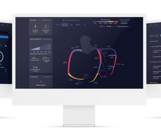
milla1cf Thu, 03/28/2024 - 07:00 March 28, 2024 — Cleerly , the company on a mission to create a new standard of care to aid in the diagnosis of heart disease, shared findings from a study published online in the Journal of the American College of Cardiology: Cardiovascular Imaging on March 13, 2024. JACC Cardiovasc Imaging.
Cardiac rupture: a clinically predictable complication of acute myocardial infarction Ventricular septal rupture == MY Comment , by K EN G RAUER, MD ( 9/5 /2024 ): == As I emphasized in My Comment in the December 6, 2022 post and the August 19, 2023 post of Dr. Smith's ECG Blog — Not all patients with acute MI report chestpain.
Circulation, Volume 150, Issue Suppl_1 , Page A4134796-A4134796, November 12, 2024. Introduction:Over 6 million patients (pts) present to US emergency departments annually with chestpain (CP), of which the majority are found to have no serious disease.
Written by Jesse McLaren A 70 year old with prior MIs and stents to LAD and RCA presented to the emergency department with 2 weeks of increasing exertional chestpain radiating to the left arm, associated with nausea. Int J Cardiol 2024 3. Eur Heart J Digital Health 2024 5. Amsterdam et al. Circulation 2014 2. Lupu et al.
Sent by anonymous A man in his 40s with no previous heart disease presented within 30 minutes of onset of acute chestpain that started while exercising. Three patients with chestpain and “normal” ECGs: which had OMI? Four patients with chestpain and ‘normal’ ECG: can you trust the computer interpretation?
We organize all of the trending information in your field so you don't have to. Join thousands of users and stay up to date on the latest articles your peers are reading.
You know about us, now we want to get to know you!
Let's personalize your content
Let's get even more personalized
We recognize your account from another site in our network, please click 'Send Email' below to continue with verifying your account and setting a password.


Let's personalize your content