This site uses cookies to improve your experience. To help us insure we adhere to various privacy regulations, please select your country/region of residence. If you do not select a country, we will assume you are from the United States. Select your Cookie Settings or view our Privacy Policy and Terms of Use.
Cookie Settings
Cookies and similar technologies are used on this website for proper function of the website, for tracking performance analytics and for marketing purposes. We and some of our third-party providers may use cookie data for various purposes. Please review the cookie settings below and choose your preference.
Used for the proper function of the website
Used for monitoring website traffic and interactions
Cookie Settings
Cookies and similar technologies are used on this website for proper function of the website, for tracking performance analytics and for marketing purposes. We and some of our third-party providers may use cookie data for various purposes. Please review the cookie settings below and choose your preference.
Strictly Necessary: Used for the proper function of the website
Performance/Analytics: Used for monitoring website traffic and interactions
The patient was discharged with a diagnosis of acute pericarditis — and treated with a full course of colchicine and ibuprofen. The ultimate discharge diagnosis was acute pericarditis. ( From the information provided — I would not make the diagnosis of acute pericarditis. Figure-1: The initial ECG in today's case.
Overall, this looks like one of the rare ECGs that is actually specific for pericarditis in my opinion. Pericarditis maybe." Meyers' words — "is one of the rare ECGs that is actually specific for pericarditis". ii ) Today's case emphasizes the importance of the history in making the diagnosis of pericarditis.
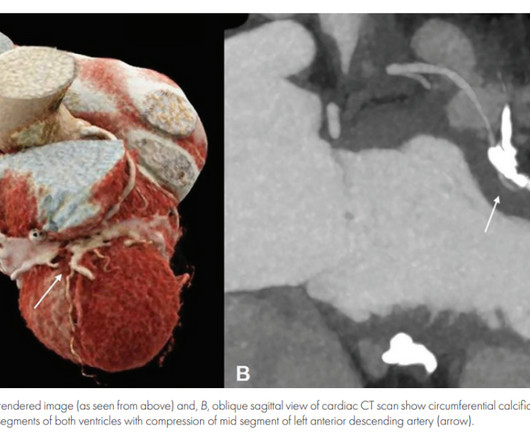
Inflammatory pericarditis can occur in differential fashion. For example, the most common chronic pericarditis tuberculosis affects the fibrinous layer. Post MI pericarditis involves the epicardium. Diastolic Coronary Artery Compression in Constrictive Pericarditis. Angina caused by calcific constrictive pericarditis.
Transition to rilonacept monotherapy from oral therapies in patients with recurrent pericarditis. Heart 2023; 109 : 297-304. Brucato A, Wheeler A, Luis SA, et al. This article has been corrected since it was first published.
This is a value typical for a large subacute MI, n ormal value 48 hours after myocardial infarction is associated with Post-Infarction Regional Pericarditis ( PIRP ). As already mentioned, this patient could have post-infarction regional pericarditis from a large completed MI. Sinus tachycardia has many potential causes. Hammill SC.
It is easy to say pericarditis in such a case. young male no risk factors and ST-elevation in several leads) As Dr. Smith has emphasized many times you diagnose pericarditis at your patient's and your own peril. Version 1 was not trained to detect myo- or pericarditis. The above ECG was recorded. How did the Queen do?
There is a reasonable chance of pericarditis in this case, or this could be a baseline." Sadly, I did not receive enough information to adjudicate whether this patient has pericarditis or not. I sent this to Dr. Smith and this was his response: "Likely pericarditis, but that is perilous. I immediately responded: "cool fake!
Recall from this post referencing this study that "reciprocal STD in aVL is highly sensitive for inferior OMI (far better than STEMI criteria) and excludes pericarditis, but is not specific for OMI." St depression in lead AVL differentiates inferior st-elevation myocardial infarction from pericarditis. link] Bischof, J. Worrall, C.,
In Europe, the TRUPULSE generator received CE mark in late 2023 and the VARIPULSE Catheter received CE mark in February 2024. In the U.S., the VARIPULSE Catheter and TRUPULSE Generator are currently investigational and are not approved by FDA. For more information: www.biosensewebster.com References: i. doi: 10.1016/j.jacc.2014.04.072
Pericarditis is rare — but myocarditis is not , so especially in this age group — more information is needed to quickly determine if this could be an acute MI, myocarditis, or none of the above. Acute coronary occlusion almost always occurs in patients who are beyond their 20s. That said — acute MI does occur in younger patients.
Circulation: Cardiovascular Imaging, Volume 16, Issue 11 , Page e015606, November 1, 2023. BACKGROUND:Pericardial late gadolinium enhancement (LGE) is usually associated with active pericarditis, but it is not infrequently found in patients after cardiac surgery even a long time after the intervention.
These latter findings are typical of pericarditis, but pericarditis never has reciprocal ST depression. Usually with pericarditis and myocarditis — hyperacute T waves (HATW) are not present. S mith : there is STE in lead III and reciprocal STD in aVL. This is OMI until proven otherwise.
First, many on Twitter said "Pericarditis". This is NOT pericarditis, which virtually NEVER has ST depression any where except aVR. See our publication: ST depression in lead aVL differentiates inferior ST-elevation myocardial infarction from pericarditis There is STE in inferior leads, high lateral leads, and V4-V6.
Of course the patient was saddled with the erroneous "pericarditis" diagnosis after CTs ruled also ruled out PE and dissection. Serial ECGs remained unchanged. Echo showed normal EF and no wall motion abnormalities, and no pericardial effusion. But he did well. 25 minutes later, EMS called back with this new ECG: Super obvious STEMI(+) OMI.
Dyspnea, Chest pain, Tachypneic, Ill appearing: Bedside Cardiac Echo gives the Diagnosis 31 Year Old Male with RUQ Pain and a History of Pericarditis. I refer to My Comment in the March 4, 2023 post — in which I included a Table with ECG Findings of Acute PE. Submitted by a Med Student, with Great Commentary on Bias!
Traditionally used as an anti-inflammatory for pericarditis (inflammation of the lining of the heart), it has recently been shown to result in fewer major heart events in those with a recent heart attack. 2023 Nov 11. It is an easy win, frequently missed. Br J Gen Pract. 2018 Mar;68(668):151-152. N Engl J Med.
PMID: 11723026. == MY Comment , by K EN G RAUER, MD ( 10/23 /2023 ): == The various forms of electrical alternans are frequently misunderstood, if not completely unnoticed. Chronic amiodarone evokes no torsade de pointes arrhythmias despite QT lengthening in an animal model of acquired long-QT syndrome. Circulation.
Bottom Line: Tests other than cardiac cath may be all that are needed to establish the diagnosis — but, I'd want to see a patient with this ECG as soon as would be possible.
Brugada Syndrome: Diagnosis and Risk Stratification Hello friends, this is the modified version of my talk at Indian Heart Rhythm Society Conference, New Delhi, 2023, on Brugada Syndrome. Hope you will enjoy this session. These are the conditions which have to be considered or excluded as they can sometimes manifest Brugada pattern on ECG.
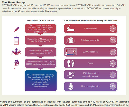
The KDCA also established a reporting system with a legal obligation for special adverse events including myocarditis and pericarditis after COVID-19 vaccination. The 2023 risk/benefit calculus casts a very long shadow on those who seek to compel their fellow citizens to vaccinate.
The "flu-like" illness suggests myo- or pericarditis, but that would be a diagnosis of exclusion. The September 15, 2023 post — for PTA ( Pulse-Tap Artifact ). The April 6, 2023 post — excessive baseline artifact misdiagnosed as AFib ( instead of sinus rhythm with AV Wenckebach — as in Figure-4 in this post ).
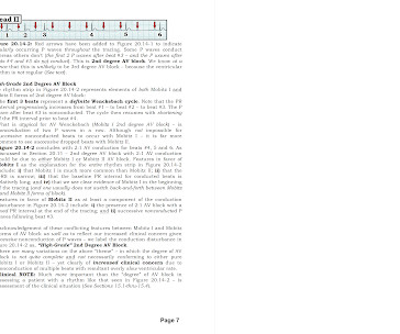
To Emphasize: When the patient is older and presents with a history of cardiac-sounding chest pain — then acute infarction will be much more common than acute pericarditis, myocarditis, or perimyocarditis. A DDENDUM ( 4/24/2023 ) : One of the most problematic areas in arrhythmia interpretation is assessment of the AV Blocks.
Pericarditis? My Comment by K EN G RAUER, MD ( 1/9 /2023 ): = “ Treat the patient — Not the age of the patient”. They still would not do angiography for her: See other cases of young patients with OMI: A 16 year old girl has syncope while playing basketball. 24 yo woman with chest pain: Is this STEMI?
False Positive ST elevation in aVL Even when the story is obvious, with intractable pain, the STEMI paradigm can cause preventable delays Man in his 60's with very subtle ECG and pain not controlled with medical therapy Pericarditis vs. MI #2 See other "Quiz Posts": Quiz post - which of these, if any, are OMI? Will you activate the cath lab?
In August, the CDC reported 29 cases of pericarditis, including five in persons with a history of pericarditis after mRNA COVID-19 vaccine ; 10 Importantly, the Novovax vaccine is a protein based vaccine that was hoped to not be associated with myocarditis as was noted with the mRNA vaccines. The pain resolved a few weeks later.
First Troponin I was <2 and peak was 8, echo showed subtle apical lateral hypokinesis, CRP was elevated, and patient was discharged with a diagnosis of regional pericarditis. In this case, there would be evolution, but the evolution would be typical of pericarditis (if the diagnosis of pericarditis was accurate!!
The initial computer and final cardiology interpretation was a differential: “ST elevation, consider early repolarization, pericarditis, or injury.” Acad Emerg Med 2023 3. Ann Emerg Med 2023 4. But STEMI criteria ignore all this and look at ST segments in isolation. Smith, Meyers. Meyers, Bracey et al.
Though less prevalent in younger patients, occlusion MI may occur and requires the same early interventions as older patients. - - Pericarditis and myocarditis should be a diagnosis of exclusion. PMID: 34013488; PMCID: PMC8134825. == MY Comment , by K EN G RAUER, MD ( 12/5 /2023 ): == Interesting case by Drs. World J Pediatr.
Prominent J waves and ventricular fibrillation caused by myocarditis and pericarditis after BNT162b2 mRNA COVID-19 vaccination. 2004 = My Comment by K EN G RAUER, MD ( 1/21 /2023 ): = I thought today's case by Drs. The relationship between J wave and ventricular tachycardia during Takotsubo cardiomyopathy.
Despite apparently hearing the above history together with two diagnostic ECGs and a troponin compatible with OMI, the cardiologist thought the ECG represented pericarditis and recommended echocardiogram. The emergency physician consulted cardiology. Several hours passed with no documentation as to the reason for delay.
Although this is not a common phenomenon You will see it on occasion ( See the June 30, 2023 post the November 27, 2023 post and the July 24, 2013 post in Dr. Smith's ECG Blog ). Smith : I recognize this as a STEMI mimic. I was not alarmed. The providers showed me the ECG and I told them that I thought it was a fake.
We organize all of the trending information in your field so you don't have to. Join thousands of users and stay up to date on the latest articles your peers are reading.
You know about us, now we want to get to know you!
Let's personalize your content
Let's get even more personalized
We recognize your account from another site in our network, please click 'Send Email' below to continue with verifying your account and setting a password.


Let's personalize your content