This site uses cookies to improve your experience. To help us insure we adhere to various privacy regulations, please select your country/region of residence. If you do not select a country, we will assume you are from the United States. Select your Cookie Settings or view our Privacy Policy and Terms of Use.
Cookie Settings
Cookies and similar technologies are used on this website for proper function of the website, for tracking performance analytics and for marketing purposes. We and some of our third-party providers may use cookie data for various purposes. Please review the cookie settings below and choose your preference.
Used for the proper function of the website
Used for monitoring website traffic and interactions
Cookie Settings
Cookies and similar technologies are used on this website for proper function of the website, for tracking performance analytics and for marketing purposes. We and some of our third-party providers may use cookie data for various purposes. Please review the cookie settings below and choose your preference.
Strictly Necessary: Used for the proper function of the website
Performance/Analytics: Used for monitoring website traffic and interactions
No ChestPain, but somnolent. The fact that this is syncope makes give it a far lower pretest probability than chestpain, but it was really more than syncope, as the patient actually underwent CPR and had hypotension on arrival of EMS. Here is the ED ECG (a photo of the paper printout) What do you think?
The patient presented due to chestpain that was typical in nature, retrosternal and radiating to the left arm and neck. He denied any exertional chestpain. It is unclear if the patient was pain free at this time. He has a medical hx notable for hypertension, hyperlipidemia and previous tobacco use disorder.
Submitted and written by Megan Lieb, DO with edits by Bracey, Smith, Meyers, and Grauer A 50-ish year old man with ICD presented to the emergency department with substernal chestpain for 3 hours prior to arrival. At this time he reported ongoing chestpain and was given aspirin and nitroglycerin.
I see the following: The rhythm is sinus bradycardia at ~55-60/minute. These tall T waves are associated with flattening ( straightening ) of the ST segment in the inferior leads — with slight S T elevation in leads V2-thru-V6 ( albeit not enough to qualify as a "STEMI" — Akbar et al, StatPearls, 2023 ).
The patient has acute chestpain. Instead — my thoughts were as follows: The rhythm is sinus , with marked bradycardia and a component of sinus arrhythmia. This was texted to me in real time. What do you think? Here was my answer: "Not ischemia. Maybe HOCM or another form of LVH. I would not activate cath lab.
The patient with no prior cardiac history presented in the middle of the night with acute chestpain, and had this ECG recorded during active pain: I did not see any ischemia on this electrocardiogram. Their apparently excessive length (QT interval) is due to bradycardia. This is a case I had quite a while back.
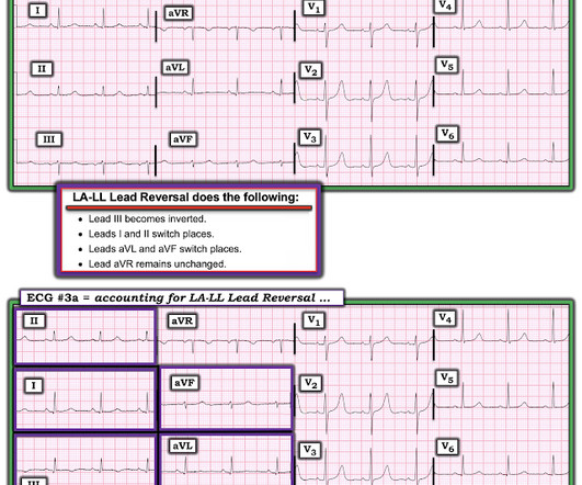
The rhythm is sinus bradycardia at a rate just over 50/minute. Although difficult to measure ( because of marked overlap of the QRS in multiple chest leads ) — there appears to be greatly increased QRS amplitude, consistent with voltage for LVH. The May 13, 2023 post ( LA-RA reversal ). All intervals ( PR,QRS,QTc ) are normal.
Written by Pendell Meyers A woman in her 50s presented with acute chestpain and lightheadedness since the past several hours. Here is her triage ECG during active symptoms: What do you think? The ED physician read this as "Normal sinus rhythm. Marked ST abnormality, possible anterior subendocardial injury."
She was hemodynamically stable — and did not have chestpain, lightheadedness or syncope. Even if we stopped here — We could conclude the following: There is marked bradycardia in today's rhythm ( ie, Heart rate in the low 30s ). QUESTIONS: HOW would you interpret the rhythm in Figure-1 ? Is this " high -grade" AV block?
Later, I found old ECGs: 5 month prior in clinic: V5 and V6 look like OMI 9 months prior in clinic with no chest symptoms: V5 and V6 look like OMI 1 year prior in the ED with chestpain: V5 and V6 sure look like a STEMI For this ECG and chestpain in the ED, the Cath lab activated. But the angiogram was clean.
to 1828 msec. ) — which corresponds to a variation in the rate of sinus bradycardia from 36-to-33/minute. This makes sense given that the underlying rhythm in today's case appears to be marked sinus bradycardia and arrhythmia , with a ventricular escape rhythm appearing when the SA node rate drops below 33/minute.
The chestpain quickly subsided. During the night, while on telemetry, the patient became bradycardic, with periods of isorhythmic AV dissociation (nodal escape rhythm alternating with sinus bradycardia), and there were sporadic PVCs. Are you worried about OMI in this case? His response was: "I would not call it OMI.
And she does not know that this is an overdose; she thinks it is a patient with chestpain!! Severely ill patients from any etiology can have very abnormal ECGs = My Comment by K EN G RAUER, MD ( 5/26 /2023 ): = There are more questions than answers in today's case. 3 hours later, this was recorded at a K of 2.8
Written by Willy Frick A 46 year old man with a history of type 2 diabetes mellitus presented to urgent care with complaint of "chest burning." The ECG shows sinus bradycardia but is otherwise normal. The patient said his chestpain was 4/10, down from 8/10 on presentation. The following ECG was obtained.
That said — obvious findings include: i ) Marked bradycardia! — A DDENDUM ( 10/28/2023 ) : This 15 -minute ECG Video ( M edia P EARL # 52) — Reviews the 3 Types of 2nd-Degree AV Block — plus — the hard-to-define term of "high-grade" AV block. The rhythm in Figure-1 is complex — and defies precise interpretation without careful study.
The combination of absence of chestpain and history of LV aneurysm made it easy to assess that this patient does not have acute OMI. In view of the History with the current admission ( ie, presenting to the ED for vertigo — with no new chestpain ) — I interpreted ECG #1 as no OMI.
Even if you don't see the OMI, you can usually prevent such a long delay to reperfusion by recording serial ECGs every 15 minutes for a patient with persistent chestpain. The rhythm in ECG #1 is sinus bradycardia and arrhythmia. Hillinger et al. The lead that immediately caught my eye — is lead aVL.
After the heart rate increased slightly, here was the repeat ECG: Sinus bradycardia, only slightly faster rate than prior. See these similar cases: A man in his sixties with chestpain Why is there inferior ST elevation, and would you get posterior leads? Sudden CP and SOB with Inferior ST Elevation and in STE in V1.
This was sent by anonymous The patient is a 55-year-old male who presented to the emergency department after approximately 3 to 4 days of intermittent central boring chestpain initially responsive to nitroglycerin, but is now more constant and not responsive to nitroglycerin. It is unknown when this pain recurred and became constant.
She did notice something slightly wrong subjectively, but had no palpitations, chestpain, or SOB, or any other symptom. Baseline bradycardia in endurance athletes limits the use of ß-blockers. This middle-aged patient has a remote history of cardiac surgery as a young child for a "heart murmur". She was on no medications.
Written by Jesse McLaren, with comments from Smith An 85 year old with a history of CAD presented with 3 hours of chestpain that feels like heartburn but that radiates to the left arm. There’s sinus bradycardia, first degree AV block, normal axis, delayed R wave progression, and normal voltages. Below is the ECG. Take home 1.
He woke up alert and with chestpain which he also had experienced intermittently over the previous few days. The history in today's case with sudden loss of consciousness followed by chestpain is very suggestive of ACS and type I ischemia as the cause of the ECG changes. What do you think?
He did not have chestpain. Chestpain in high risk patient. Syncope and Bradycardia Syncope in a 20-something woman Long QT: Do not trust the computerized QT interval when the QT is long An Alcoholic Patient with Syncope Cardiac Arrest. Here is his triage ECG: What do you think? Is it STEMI? What is going on here?
All of the patients presented with chestpain , and they are all in triage. Triage is backed up, and 10 minutes into your shift one of the ED nurses brings your several ECG s that has not been overread by a physician. Which, if any, of these patients has OMI, with myocardium at risk and need for emergent PCI?
There was no chestpain. For example — bradycardia and AV conduction disturbances are not uncommon with Hyperkalemia , with these conduction disturbances most often resolving once serum K+ is corrected. This was written by Magnus Nossen The patient is a female in her 50s. She was feeling fine prior to the last seven days.
To improve visualization — I've digitized the original ECG using PMcardio ) MY Thoughts on the ECG in Figure-1: This is a challenging tracing to interpret — because there is marked bradycardia with an irregular rhythm and a change in QRS morphology. Figure-1: The initial ECG in today's case. ( The QRS complex is wide ( ie, >0.10
Patient 2 : 55 year old with 5 hours of chestpain radiating to the shoulder, with nausea and shortness of breath ECG: sinus bradycardia, normal conduction, normal axis, normal R wave progression, no hypertrophy. This was missed by the treating physician, but the chestpain resolved with aspirin.
A man in his early 30s was walking when he developed central chestpain which was non-radiating, then had a syncopal event with bowel incontinence, and when he woke up he had ongoing chestpain. Notes never having symptoms like this before, pain is so severe its causing SOB. He called 911. As I wrote in that Nov.
days of chestpain that started as substernal and crushing in nature awakening him from sleep and occasionally traveling to right side of neck. The pain was described as constant, worse with deep inspiration and physical activity, sometimes sharp. He reported 1.5 World J Pediatr. 2021 Aug;17(4):335-340. Epub 2021 May 20.
Within ten minutes, she developed bradycardia, hypotension, and ST changes on monitor. Bradycardia and heart block are very common in RCA OMI. He had no chestpain, dyspnea, or any other anginal equivalent, and his vital signs were normal. He told the patient this horrible news. European Heart Journal , 39 (22), 20322046.
We organize all of the trending information in your field so you don't have to. Join thousands of users and stay up to date on the latest articles your peers are reading.
You know about us, now we want to get to know you!
Let's personalize your content
Let's get even more personalized
We recognize your account from another site in our network, please click 'Send Email' below to continue with verifying your account and setting a password.


Let's personalize your content