This site uses cookies to improve your experience. To help us insure we adhere to various privacy regulations, please select your country/region of residence. If you do not select a country, we will assume you are from the United States. Select your Cookie Settings or view our Privacy Policy and Terms of Use.
Cookie Settings
Cookies and similar technologies are used on this website for proper function of the website, for tracking performance analytics and for marketing purposes. We and some of our third-party providers may use cookie data for various purposes. Please review the cookie settings below and choose your preference.
Used for the proper function of the website
Used for monitoring website traffic and interactions
Cookie Settings
Cookies and similar technologies are used on this website for proper function of the website, for tracking performance analytics and for marketing purposes. We and some of our third-party providers may use cookie data for various purposes. Please review the cookie settings below and choose your preference.
Strictly Necessary: Used for the proper function of the website
Performance/Analytics: Used for monitoring website traffic and interactions
= Case Presentation by K EN G RAUER, MD ( 5/5 /2023 ): — Edits by Drs. The "good" news — Treatment with naloxone will probably resolve the bradycardia. Meyers & Smith. = Dr. Smith was reading ECGs — and he sent myself and Dr. Meyers the tracing shown in Figure-1. At the time we did not yet know the history. What do YOU think?
Discontinue all negative chronotropic agents, since the risk of torsade is much higher with bradycardia or pauses. As described above by Dr. Smith Pacing in today's case is an effective intervention as doing so prevents the bradycardia and pauses that are likely to precipitate additional episodes of Torsades de Pointes. (
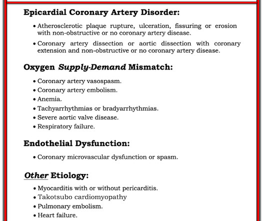
As per my review of this subject ( Check out My Comment at the bottom of the page in the November 16, 2023 post in Dr. Smith's ECG Blog ) — the 3 most common Causes of ACS ( A cute C oronary S yndrome ) with a "negative" cath are: i ) Myocarditis; ii ) Takotsubo cardiomyopathy; and , iii ) MINOCA.
The primary outcome was ventricular arrhythmias, the secondary outcomes were bradycardia and atrial fibrillation (AF).ResultsOur 0.66], but increased the risk of in-hospital bradycardia (OR 2.88, 95% CI 1.02–8.17) 8.17) compared with propofol.
Altered Mental Status, Bradycardia == MY Comment , by K EN G RAUER, MD ( 2/2 /2024 ): == Dr. Meyers began today’s case with the clinical challenge of asking you to identify the underlying cause of ECG #2. -- Read this ECG -- Osborn Waves and Hypothermia (this is the "Figure" above) What does LBBB look like in severe hypothermia?
to 1828 msec. ) — which corresponds to a variation in the rate of sinus bradycardia from 36-to-33/minute. This makes sense given that the underlying rhythm in today's case appears to be marked sinus bradycardia and arrhythmia , with a ventricular escape rhythm appearing when the SA node rate drops below 33/minute.
Even if we stopped here — We could conclude the following: There is marked bradycardia in today's rhythm ( ie, Heart rate in the low 30s ). Finally — If today's patient does not have significant underlying coronary disease — then her bradycardia with AV block may be the result of SSS ( S ick S inus S yndrome ).
The rhythm is sinus bradycardia at a rate just over 50/minute. The May 13, 2023 post ( LA-RA reversal ). The May 30, 2023 post ( LA-RA reversal ). Figure-2: The repeat ECG in today's case — recorded as soon as the treating clinician recognized the lead misplacement. ( All intervals ( PR,QRS,QTc ) are normal.
I did not think that the T-waves in V2 and V3 are hyperacute and I still do not--I disagree with Ken below--I think they are normal , especially in the context of bradycardia. Their apparently excessive length (QT interval) is due to bradycardia. They do not have much bulk. A corrected QT would be normal.
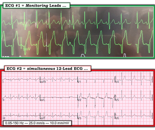
During the night, while on telemetry, the patient became bradycardic, with periods of isorhythmic AV dissociation (nodal escape rhythm alternating with sinus bradycardia), and there were sporadic PVCs. This is what T-waves look like when there is a long QT." Below are two ECGs from the telemetry monitoring.
I see the following: The rhythm is sinus bradycardia at ~55-60/minute. These tall T waves are associated with flattening ( straightening ) of the ST segment in the inferior leads — with slight S T elevation in leads V2-thru-V6 ( albeit not enough to qualify as a "STEMI" — Akbar et al, StatPearls, 2023 ).
Acute MI per se usually does not depress cardiac function and blood pressure enough to cause syncope ( Mostafa et al — J Com Hosp Intern Med Perspect 13(4):9-12, 2023 - ). Other cardiac-related causes for syncope associated with acute MI may include malignant ventricular arrhythmias and bradyarrhythmias including AV block.
Methods The TriNetX database (Jan 2015June 2023) was queried to include adults receiving their first single-chamber ventricular pacemaker for heart block, bradycardia, sick sinus syndrome, atrial fibrillation, or syncope, excluding those with prior pacemakers, heart failure or enrolled for research settings.
link] == MY Comment , by K EN G RAUER, MD ( 11/11 /2023 ): == From a learning standpoint — I LOVED this case because it illustrates in many ways how some patients simply "do not read the textbook before they come to the ED". It is not yet available, but this is your way to get on the list.
ABSTRACT Introduction Leadless pacemakers (LPs) are a valuable treatment for bradycardia, with the Aveir offering advanced features, including a protective sleeve and active fixation. The higher impedance during the implantation was a strong predictor of a midterm favorable PCT. A PCT elevation was defined as that >1.5V/0.4ms
Severely ill patients from any etiology can have very abnormal ECGs = My Comment by K EN G RAUER, MD ( 5/26 /2023 ): = There are more questions than answers in today's case. If the situation is not right for acute coronary occlusion, then the ECG findings probably do not represent acute coronary occlusion.
That said — obvious findings include: i ) Marked bradycardia! — A DDENDUM ( 10/28/2023 ) : This 15 -minute ECG Video ( M edia P EARL # 52) — Reviews the 3 Types of 2nd-Degree AV Block — plus — the hard-to-define term of "high-grade" AV block. The rhythm in Figure-1 is complex — and defies precise interpretation without careful study.
The August 17, 2020 post by me in Dr. Smith's ECG Blog — in which I review the phenomenon of Bradycardia-dependent BBB ( sometimes called "Phase 4" or "paradoxical" block ). A DDENDUM ( 12/23/2023 ) : I've presented this material before — but it bears repeating for reference. ECG Blog #242 — Reviews rate -related BBB.
He suffered from symptomatic bradycardia and falls for over a month and was seen by another cardiologist specifically for presyncope, and still the problem went unrecognized despite ongoing ECG evidence of pacemaker malfunction, and imaging proving lead dislodgement.
The ECG shows sinus bradycardia but is otherwise normal. Written by Willy Frick A 46 year old man with a history of type 2 diabetes mellitus presented to urgent care with complaint of "chest burning." The documentation does not describe any additional details of the history. The following ECG was obtained. ECG 1 What do you think?
For example — marked bradycardia with unusual forms of advanced AV block — or marked bradycardia without evident P waves — or marked QRS widening with "shapeless" morphology — are all ECG indication of the need for immediate IV calcium until improvement of these ECG patterns.
After the heart rate increased slightly, here was the repeat ECG: Sinus bradycardia, only slightly faster rate than prior. Hint: there are 2 etiologies) = Comment by K EN G RAUER, MD ( 2/23 /2023 ): = As today's case was contributed by Dr. Nossen, who practices in Norway — today's ECG uses the Cabrera Format.
Instead — my thoughts were as follows: The rhythm is sinus , with marked bradycardia and a component of sinus arrhythmia. In this case, QOH was incorrect , saying "OMI High confidence." Smith's — in that despite the alarming ST-T wave changes, I did not think ECG #1 was the result of an acute event.
I have periodically called attention to examples of the Ashman phenomenon as they occur in Dr. Smith's ECG Blog ( See My Comments in the January 5, 2020 post — the June 17, 2020 post — and the March 30, 2023 post , among others ).
I sent this ECG to the Queen of Hearts (PMcardio OMI), and here is the verdict: == MY Comment , by K EN G RAUER, MD ( 7/26 /2023 ): == As regular followers of Dr. Smith’s ECG Blog have seen — some of the best cases that we post come from tracings that Dr. Smith or Meyers see while simply checking out “tracings on the system”.
Triage physician interpretation: -sinus bradycardia -lateral ST depressions While there are lateral ST depressions (V5, V6) the deepest ST depressions are in V4. My Comment by K EN G RAUER, MD ( 1/23 /2023 ): = The importance of today's case by Drs. Triage EKG: What do you think?
My Comment by K EN G RAUER, MD ( 3/15 /2023 ): = I found today’s case highly instructive in highlighting a number of important aspects regarding the presentation and initial treatment of a patient who presents to the ED with new AFib. Baseline bradycardia in endurance athletes limits the use of ß-blockers.
The patient later settled into sinus bradycardia. The amiodarone was discontinued and the patient did well. == MY Comment , by K EN G RAUER, MD ( 6/23 /2023 ): == From an academic standpoint — I love WCT ( W ide- C omplex T achycardia ) rhythms.
My Comment by K EN G RAUER, MD ( 1/15 /2023 ): = For many reasons — I thought today's post by Dr. Smith to be highly insightful and extremely useful to any provider charged with interpreting emergency ECGs. The rhythm in ECG #1 is sinus bradycardia and arrhythmia. Hillinger et al. The lead that immediately caught my eye — is lead aVL.
The RCA usually supplies the SA and AV nodes, so RCA OMIs can present with heart blocks and bradycardias like in this case. == MY Comment , by K EN G RAUER, MD ( 7/30 /2023 ): == There are numerous important points that are brought out by today’s case. Subendocardial ischemia does not localize.
milla1cf Fri, 01/19/2024 - 10:55 January 19, 2024 — Orchestra BioMed , a biomedical company accelerating high-impact technologies to patients through risk-reward sharing partnerships, today announced the first patient was randomized in the BACKBEAT pivotal study in late December 2023. Kowal, M.D.,
For example — bradycardia and AV conduction disturbances are not uncommon with Hyperkalemia , with these conduction disturbances most often resolving once serum K+ is corrected. A DDENDUM: There are many potential causes of AV block.
As per Dr. Nossen — today's initial ECG ( LEFT tracing in Figure-2 ) shows sinus bradycardia with QRS widening due to bifascicular block ( RBBB/LAHB ). And although severe underlying coronary disease is often the cause — non-coronary causes may be seen ( See My Comment in the March 1, 2023 post for the common causes of DSI ).
That is what Emergency Medicine is all about. == MY Comment , by K EN G RAUER, MD ( 9/3 /2023 ): == I’ve seen a “common theme” with many of the ECGs that Drs. MY Thoughts on the ECG in Figure-1: The rhythm in ECG #1 is sinus bradycardia at ~50-55/minute. Figure-1: The initial ECG in today's case. Intervals ( PR, QRS, QTc ) are normal.
There’s sinus bradycardia, first degree AV block, normal axis, delayed R wave progression, and normal voltages. Hyperacute T waves are deflating, suggesting reperfusion but there is still reciprocal change in I/aVL and ST depression in V2, and the bradycardia is worse. Below is the ECG. What do you think? Take home 1.
The rhythm is uncertain ( ie, We only see 4 beats — because the same 4 beats are repeated in limb and chest leads — but in lead II there appears to be sinus bradycardia and arrhythmia plus a P wave with a PR interval too short to conduct preceding beat #1 — therefore need for a longer period of monitoring ).
Syncope and Bradycardia Syncope in a 20-something woman Long QT: Do not trust the computerized QT interval when the QT is long An Alcoholic Patient with Syncope Cardiac Arrest. Another diagnostic ECG of a potentially deadly condition == MY Comment , by K EN G RAUER, MD ( 10/19 /2023 ): == Today's excellent case discussion by Drs.
Stroke: Vascular and Interventional Neurology, Volume 3, Issue S2 , November 1, 2023. Six patients (17.1%) experienced perioperative complications including stent occlusion, bradycardia, and hematoma. IntroductionIntracranial atherosclerosis (ICAS) is a leading global cause of stroke.
To improve visualization — I've digitized the original ECG using PMcardio ) MY Thoughts on the ECG in Figure-1: This is a challenging tracing to interpret — because there is marked bradycardia with an irregular rhythm and a change in QRS morphology. Figure-1: The initial ECG in today's case. ( The QRS complex is wide ( ie, >0.10
Theres sinus bradycardia, borderline PR interval, narrow QRS; normal axis/R wave progression; low precordial voltages, and subtle peaked T waves (most obvious in V2, but all T waves are symmetric with a narrow base). Theres no prior ECG to compare - but the bradycardia, prolonged PR and peaked T waves could all be from hyperkalemia.
Patient 2 : 55 year old with 5 hours of chest pain radiating to the shoulder, with nausea and shortness of breath ECG: sinus bradycardia, normal conduction, normal axis, normal R wave progression, no hypertrophy. Smith : The fact that the ECG did not evolve is further proof that this was the baseline ECG. nearly identical to the first case).
There is also STE in V1 which is diagnostic of right ventricular OMI in this situation , and partly explains the syncope and hypotension (along with the bradycardia). As I wrote in My Comment in the November 16, 2023 post in Dr. Smith's ECG Blog "I suspect that the entity known as MINOCA is not fully appreciated by many clinicians."
As I emphasized in My Comment in the February 27, 2023 post in Dr. Smith's ECG Blog some patients may develop everything except QRS widening. Among the "lessons" from today's case are the following: Although many patients follow the "textbook sequence" of ECG changes with their hyperkalemia a significant percentage of patients do not.
2023 Sep 29;15(9). Simple measures, optimal BP, like weight reduction, (Atrial interstitial fat shedding) , relaxation can prevent 90% of AF burden. Ahammed MR et al Impact of Weight Loss on Atrial Fibrillation. Regarding pharmaco-therapy, the celebrated vintage days of anti-arrhythmic drugs have almost gone.
We organize all of the trending information in your field so you don't have to. Join thousands of users and stay up to date on the latest articles your peers are reading.
You know about us, now we want to get to know you!
Let's personalize your content
Let's get even more personalized
We recognize your account from another site in our network, please click 'Send Email' below to continue with verifying your account and setting a password.


Let's personalize your content