This site uses cookies to improve your experience. To help us insure we adhere to various privacy regulations, please select your country/region of residence. If you do not select a country, we will assume you are from the United States. Select your Cookie Settings or view our Privacy Policy and Terms of Use.
Cookie Settings
Cookies and similar technologies are used on this website for proper function of the website, for tracking performance analytics and for marketing purposes. We and some of our third-party providers may use cookie data for various purposes. Please review the cookie settings below and choose your preference.
Used for the proper function of the website
Used for monitoring website traffic and interactions
Cookie Settings
Cookies and similar technologies are used on this website for proper function of the website, for tracking performance analytics and for marketing purposes. We and some of our third-party providers may use cookie data for various purposes. Please review the cookie settings below and choose your preference.
Strictly Necessary: Used for the proper function of the website
Performance/Analytics: Used for monitoring website traffic and interactions
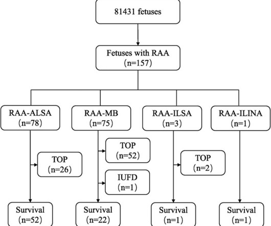
RAA features were characterised by comparing prenatal ultrasound data with anatomical casting results after pregnancy termination or postnatal imaging and surgical intervention to analyse the prognosis and misdiagnoses of fetal RAA.ResultsOf the 157 fetal RAA cases, 50 (31.8%) cases were isolated RAA and 107 (68.2%) cases were nonisolated RAA.
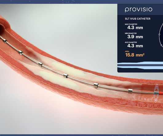
The SLT IVUS Support Crossing Catheter is an over-the-wire intravascular ultrasound catheter with an ultrasound transducer array at the distal end that also functions as a support crossing catheter. Intravascular Ultrasound Imaging Versus Digital Subtraction Angiography in Patients with Peripheral Vascular Disease.
Let us explore how Smart Cath Labs can be advanced in the year 2022 Here we have explored five innovations for new Cath Lab technology, including predictive analytics, augmenting soft-tissue visualisation, robotics, trimming down the usage of X-ray navigation and Digital Cath Lab solutions. The post How to Build Smart Cath Labs in 2022?
The algorithm uses deep learning to analyse routine ultrasound scans of the heart ( echocardiograms ) to detect disease that often goes undetected during standard assessments.
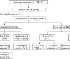
Objective The study aims to assess the ultrasonic features of fetal cardiac rhabdomyoma (CR), track the perinatal outcome and postnatal disease progression, investigate the clinical utility of ultrasound, MRI and tuberous sclerosis complex (TSC) gene analysis in CR evaluation, and offer evidence for determing of fetal CR prognosis.
The incidence of no-reflow was higher in patients with attenuated plaque ≥5 mm in length as evaluated by intravascular ultrasound (IVUS).Objective:The The incidence of no-reflow was higher in patients with attenuated plaque ≥5 mm in length as evaluated by intravascular ultrasound (IVUS).Objective:The
Answer : Bedside ultrasound! Smith : RV infarct may also have this appearance on ultrasound. So hypoxia without B lines on lung ultrasound strongly weights toward PE. What do you do clinically when the ECG looks like this? How do you emergently triage the patient to cath lab or possibly IR suite for thrombectomy?
Few of them currently have the equipment and expertise to diagnose valvular heart disease, but recent studies have demonstrated that healthcare professionals can use a combination of portable ultrasound devices and AI to diagnose heart diseases as well.
Bedside cardiac ultrasound with no obvious wall motion abnormalities. Review of the 2 ECGs in today's case is insightful ( Figure-1 ): The initial ECG shows sinus rhythm, LAHB and meets Peguero Criteria for LVH ( See My Comment in the August 15, 2022 post of Dr. Smith's ECG Blog for more on LVH criteria ). He was started on nitro gtt.
However, IVI is underutilized and is not yet established as a performance measure for quality PCI.METHODS:We examined temporal trends of IVI use for all PCIs performed at Veterans Affairs hospitals in the United States from 2010 to 2022 using retrospective observational cohorts. in 2022 in 136 071 PCIs included in the study.
He arrived in the ED and had an immediate bedside cardiac ultrasound while this ECG was being recorded. The bedside ultrasound (video not available) reportedly showed only a slightly reduced LV function. The patient was given 6mg, then 12 mg, of adenosine, without a change in the rhythm. Here is the ECG: What do you think?
Methods We conducted a retrospective analysis of 121 prenatally diagnosed patients with TGA at our hospital between January 2012 and September 2022. This analysis included prenatal ultrasound, prenatal screening, clinical management and follow-up procedures.
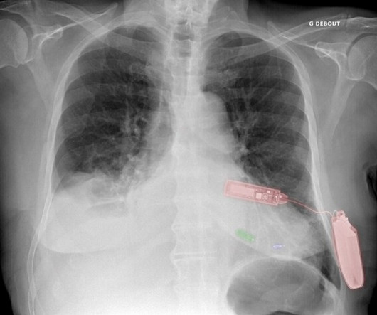
Green: Micra leadless pacemaker; blue: WiSE-CRT system LV endocardial electrode; and red: WiSE-CRT system subcutaneous battery and ultrasound generator. Leadless Ultrasound-Based Cardiac Resynchronization System in Heart Failure. 2022 Jan;19(1):22-29. A patient chest X-ray showing both Micra and WiSE-CRT systems. Reference 1.
Bedside ultrasound with no apparent wall motion abnormalities, no pericardial effusion, no right heart strain. Despite description of Wellens’ Syndrome over 40 years ago — this syndrome remains misunderstood by all-to-many clinicians ( See My Comment at the bottom of the page in the August 12, 2022 post in Dr. Smith’s ECG Blog ).
Background:Since January 2022, our emergency medical center has operated a prehospital extracorporeal cardiopulmonary resuscitation (ECPR) system. Cannulation is performed in a mobile ICU under mechanical CPR, with ultrasound-guided cannulation as the first choice. The ECPR team is dispatched for suspected CPA cases.
Carotid ultrasound results were divided into two groups based on the presence or absence of plaque. Lp(a) levels were categorized into two groups: below 50 mg/dl and 50 mg/dl or higher. Coronary artery calcium score CT results were analyzed in two groups: CACS=0 and CACS>0.Results:A Results:A total of 2620 subjects were enrolled.
24: Joint American College of Cardiology/Journal of the American College of Cardiology Late-Breaking Clinical Trials (Session 402) Saturday, April 6 9:30 – 10:30 a.m.
Methods Using endoscopy, endoscopic ultrasound, and electrogastrography before and after PVI, esophageal and periesophageal injury (mucosal lesions, food retention, periesophageal edema, or vagal nerve injury) were assessed following PFA and radiofrequency (RF)- or cryoballoon (CB)-PVI.
This study aimed to determine the current prevalence of CAS and examine the associated gender differences in adults.Methods:From September 2021 to June 2022, we established a prospective cohort to study CAS and cardiovascular disease across 25 project sites in Henan, China, utilizing a multistage whole-population sampling method.
A majority (62.5%) of those presenting with ‘normal’ ECGs had the cath lab activated without any ECG being labeled ‘STEMI’ by automated interpretation – based on signs of Occlusion MI including ECG changes, regional wall motion abnormality on bedside ultrasound, or refractory ischemia. 2022 ; 51 : 384 - 387 3. 2022 ; 55 : 180 - 182 6.
The primary efficacy outcome was the composite of no occlusion in the treated segment assessed at serial duplex ultrasound examinations or no reintervention needed to maintain patency within 6 months. Secondary outcomes, including Villalta score, quality of life, and safety outcomes, were also assessed.
Case continued A bedside ultrasound showed diminished LV EF and of course bradycardia. The April 17, 2022 post ( Leads V1,V2 misplacement ). The May 24, 2022 post ( LA-LL reversal ). The May 26, 2022 post ( LA-LL reversal ). The August 17, 2022 post ( LA-RA reversal ). RVMI explains part of the shock.
2 High intensity interval training induces beneficial effects on coronary atheromatous plaques – a randomized trial, European Journal of Preventive Cardiology , 2022;, zwac309, 3 The effects of lipid-lowering therapy on coronary plaque regression: a systematic review and meta-analysis. Springer, Cham. Sci Rep 11 , 7999 (2021).
Pads were placed with ultrasound guidance, so they were in the correct position. As I discussed and documented in Lesson 1 of My Comment at the bottom of the page in the April 2, 2022 post of Dr. Smith's ECG Blog — certain patients may remain in sustained VT not only for hours — but even for days! However, this is not SVT.
All patients had standard TTE and an rTCD (NovaSignal) with saline bubble injection at rest and Valsalva strain for assessment of RLS performed by echo and ultrasound technicians. Average age was 55.7 ± 11.1
It was notable for a normal cardiac ultrasound with no pericardial fluid, normal LV and RV function (though the quality was not sufficient to evaluate for wall motion abnormalities) and normal IVC dynamics. Bedside ultrasound is another very important piece. Ultrasound can be very helpful to distinguish causes of hypotension.
Fortunately, this operator used intravascular ultrasound (IVUS). NOTE: For review of 20 cases of "Swirl" vs Swirl "Look-Alikes" — Check out the October 15, 2022 post in Dr. Smith's ECG Blog. An angiogram is a " lumenogram " and does not "see" the extraluminal plaque. Most plaque is outside the lumen!!
Cardiac Ultrasound may be a surprisingly easy way to help make the diagnosis Answer: pulmonary embolism. Now another, with ultrasound. NOTE: For more on the ECG diagnosis of acute RV "strain" ( and acute PE ) — Please check out My Comment at the bottom of the page in the March 28, 2022 post in Dr. Smith's ECG Blog.
We report our experience validating this large animal model for translational and preclinical research.Methods3 animal experiments were performed at the UCLA TRIC laboratory between 02/2022 and 10/2022. The sheep were anesthesitized and handled under approval of the Animal Research Committee at UCLA (protocol ARC 2020‐19).
This case was provided by Spencer Schwartz, an outstanding paramedic at Hennepin EMS who is on Hennepin EMS's specialized "P3" team, a team that receives extra training in advanced procedures such as RSI, thoracostomy, vasopressors, and prehospital ultrasound. Takotsubo is a sudden event, not one with crescendo angina.
Another approach is sympathetic chain (stellate ganglion) blockade if you have the skills to do it: it requires some expertise and ultrasound guidance. Meyers and Smith illustrate 20 example cases vs "look-alikes" of Swirl ( with my synthesis of "Swirl" ECG findings in My Comment on that post ) from October 15, 2022.
The problem is difficult to study because angiographic visualization of arteries is not perfect, and not all angiograms employ intravascular ultrasound (IVUS) to assess for unseen plaque or for plaque whose rupture and ulceration cannot be seen on angiogram. Thus, intracoronary imaging modalities are crucial in this setting.
Regional wall motion abnormality-inferolateral (this is the formal ultrasound location of a posterior wall motion abnormality). 2022 ACC expert consensus decision pathway on the evaluation and disposition of acute chest pain in the emergency department: A report of the American college of cardiology solution set oversight committee. •
I would do bedside ultrasound to look at the RV, look for B lines as a cause of hypoxia (which would support OMI, and argue against PE), and if any doubt persists, a rapid CT pulmonary angiogram. As for the ECG, it could represent OMI, but RBBB is also a clue that it may be PE.
No pericardial effusion on ultrasound." We've previously discussed the all-too-often ignored entity known as MINOCA ( = MI with N on- O bstructive C oronary A rteries ) — which we detailed in the November 30, 2022 post in Dr. Smith's ECG Blog ( See My Comment at the bottom of the page ). What do you think?
Dr. Nossen performed a bedside ultrasound which was interpreted as normal. For readers not familiar with this format — I've reviewed its features in the Addendum at the bottom of the page in the April 24, 2022 post in Dr. Smith's ECG Blog. Nossen, who practices in Norway — today's ECG uses the Cabrera Format.
My bedside ultrasound was of insufficient quality, but showed somewhat reduced overall EF, distended IVC without respiratory variation, no pericardial effusion, and diffuse bilateral B lines. == What do you think of her ECG? Please see My Comment in the May 14, 2022 post for a regular WCT that turned out to be antidromic AVRT.
You use an ultrasound. Nat Rev Cardiol 19 , 168–179 (2022). Regardless of the murmur findings they describe. Because using the sounds of a murmur you hear with a stethoscope that was invented in the 1700s is NOT how you make a diagnosis today. Which can now be used easily at the bedside. i.e. You DO NOT GUESS! Shapiro, M.D.
A lower extremity arterial ultrasound revealed elevated velocities in the right proximal superficial femoral artery. A second opinion from another physician at another facility resulted in a normal ultrasound. The resulting investigation and sanction to suspend his medical license did not take place until late in 2022.
Routine STEMI activation in STE-aVR for emergent revascularization is not warranted, although urgent, rather than emergent, catheterization appears to be important. == MY Comment, by K EN G RAUER, MD ( 11/4 /2022 ): == Our thanks to Drs. A emergent cardiology consult can be helpful for equivocal cases.
A bedside cardiac ultrasound was performed with a parasternal long axis view demonstrated below: There is a large pericardial effusion with collapse of the right ventricle during systole. The beat-to-beat variation in QRS amplitude and morphology is electrical alternans. This patient is only pseudo-stable. She has already had syncope.
This latest data dump did not arrive soon enough to affect the ACIP committee’s recommendations but should serve to chill the ambition of exuberant, overconfident bureaucrats still seeking to mandate vaccines in the fall of 2022. That work is far from complete. Anish Koka is a cardiologist.
Clin Cardiol 2022; [link] Labs included: hsTnI 156 ng/L, Hb 12 g/dL, WBC 12x10^9/L, Cr. Smith comment: Point of Care ultrasound is not adequate to rule out wall motion abnormality; moreover, diffuse subendocardial ischemia often has no wall motion abnormality because the epicardium is still contracting. Lupu L, et al. mg/dL, K 3.5
We organize all of the trending information in your field so you don't have to. Join thousands of users and stay up to date on the latest articles your peers are reading.
You know about us, now we want to get to know you!
Let's personalize your content
Let's get even more personalized
We recognize your account from another site in our network, please click 'Send Email' below to continue with verifying your account and setting a password.


Let's personalize your content