This site uses cookies to improve your experience. To help us insure we adhere to various privacy regulations, please select your country/region of residence. If you do not select a country, we will assume you are from the United States. Select your Cookie Settings or view our Privacy Policy and Terms of Use.
Cookie Settings
Cookies and similar technologies are used on this website for proper function of the website, for tracking performance analytics and for marketing purposes. We and some of our third-party providers may use cookie data for various purposes. Please review the cookie settings below and choose your preference.
Used for the proper function of the website
Used for monitoring website traffic and interactions
Cookie Settings
Cookies and similar technologies are used on this website for proper function of the website, for tracking performance analytics and for marketing purposes. We and some of our third-party providers may use cookie data for various purposes. Please review the cookie settings below and choose your preference.
Strictly Necessary: Used for the proper function of the website
Performance/Analytics: Used for monitoring website traffic and interactions
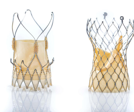
IntroductionSince TAVR was approved for lower-risk aortic stenosis (AS) patients, managing post-implantation conduction disturbances has become crucial, especially with self-expanding heart valves (SEV).
BackgroundOur previous preclinical study demonstrated thatAPOE4targeted replacement mice exhibit more severe cerebral hypoperfusion and cognitive impairment thanAPOE3targeted replacement mice with carotid artery stenosis due to neurovascular dysfunction.
Thus, this study is to observe the association between MIC and cardiac function in patients with CHD.MethodsAll participants were recruited from the Department of Cardiology, Peking University People's Hospital from August 2022 to September 2023. Among them, a total of 39 patients diagnosed with MIC and CHD were enrolled in the MIC-CHD group.
Since the pathologist does not know the original cross-sectional area of the artery or the amount of compensatory enlargement of the artery from evaluation of a single cross section of the artery at a site of stenosis, the degree of luminal narrowing of that segment cannot be determined. These are typical findings at sites of plaque rupture.
Baseline angiograms were reviewed to assess the presence or absence of intracranial stenosis lesions (IS+ Vs IS-) different than the target occlusion. Further research is warranted to explore the diagnostic value of multiple intracranial stenosis in patients undergoing mechanical thrombectomy. minutes; p=0.018). Vs 3; p=0.036).Conclusions:Our
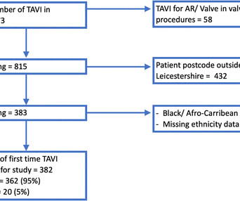
Objectives To explore the ethnic differences in patients undergoing aortic valve (AV) intervention for severe aortic stenosis (AS) in Leicestershire, UK. Results Of the 1231 SAVR and 815 TAVI performed, 6.5% were in ethnic minority patients, respectively.
Background: Symptomatic severe aortic stenosis (AS) remains undertreated with high resultant mortality despite increased growth and availability of aortic valve replacement (AVR) since the advent of transcatheter therapies. Circulation, Ahead of Print. cm2were enrolled. cm2were enrolled.
2022 Aug, 80 (9) 934946 Kronenberg F. et al, Lipoprotein(a) in atherosclerotic cardiovascular disease and aortic stenosis: a European Atherosclerosis Society consensus statement , European Heart Journal, Volume 43, Issue 39, 14 October 2022, Pages 39253946 Koschinsky ML et al.
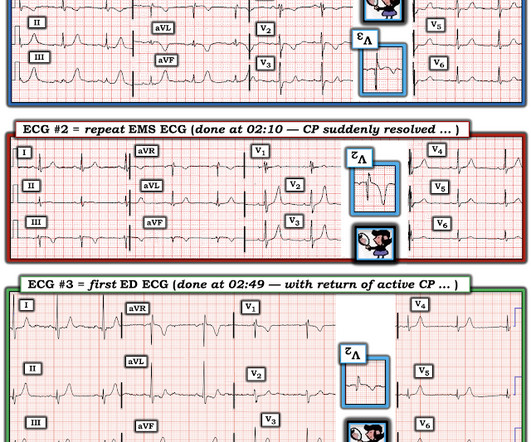
BUT — Cardiac catheterization done a little later did not reveal any significant stenosis. Despite the absence of significant coronary stenosis on her post-arrest cath — the ECG in Figure-1 is clearly diagnostic of an extensive anterolateral STEMI ( presumably from acute LAD [ L eft A nterior D escending ] coronary artery occlusion).
Despite description of Wellens’ Syndrome over 40 years ago — this syndrome remains misunderstood by all-to-many clinicians ( See My Comment at the bottom of the page in the August 12, 2022 post in Dr. Smith’s ECG Blog ). Angiography : --Culprit for the patient's unstable angina/Wellen syndrome is a ruptured plaque in the mid LAD. --As
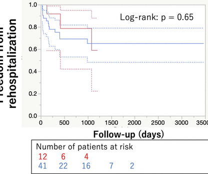
BACKGROUND:This study aimed to compare the incidence and prognostic implications of new-onset conduction disturbances after surgical aortic valve replacement (SAVR) in patients with bicuspid aortic valve (BAV) aortic stenosis (AS) versus patients with tricuspid aortic valve (TAV) AS (ie, BAV-AS and TAV-AS, respectively).
Materials and methods This retrospective, institutional-review board approved study included consecutive patients with calcified coronary artery plaques undergoing CCTA with PCD-CT and invasive coronary angiography between July and December 2022. Virtual monoenergetic images (VMI) and VNCa images were reconstructed. 68, ICC: 94 and 1%, p = .07,
Coronary CTA With AI-QCT Interpretation: Comparison With Myocardial Perfusion Imaging for Detection of Obstructive Stenosis Using Invasive Angiography as Reference Standard. AJR Am J Roentgenol 2022 Sep;219(3):407-419. Journal of Cardiovascular Computed Tomography Vol. 7 Bär S, Nabeta T, Maaniitty T.
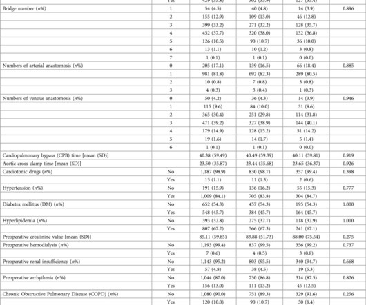
Methods We retrospectively analysed data from 745 consecutive patients who underwent TAVI for severe aortic stenosis from November 2013 to July 2022. To seek the predictors and clinical impacts of PPMI and investigate the recovery rate from conduction disorders. Results Postoperative PPMI was performed in 7.1% (53/745) of patients.
Percentage of stenosis was 50% (IQR, 36%58%). mm (IQR, 7.59.5) (P<0.001), with median stenosis expansion at 103% (IQR, 51%146%). ConclusionsOptimusL stents can safely treat arterial and venous stenosis in infants and small children via a lowprofile approach with good outcomes. male) with median age and weight of 3.4
However, it has not been clinically investigated in patients with asymptomatic CASO.Methods:A cross-sectional observational study was conducted between January 2017 and March 2022.
However, the association between the extent of cardiac damage at the time of AVR and health care costs and resource utilization has never been described.METHODS:The Optum de-identified Market Clarity database was used to identify patients with aortic stenosis treated with AVR between 2016 and 2022.
Even in patients whose moderate stenosis undergoes thrombosis, most angiograms show greater than 50% stenosis after the event. However, one can certainly imagine that many thromboses of non-obstructive lesions completely lyse and do not leave a stenosis on same day or next day angiogram.
33 preoperative clinical features and 4 postoperative complications were collected in each group. LASSO is a regression analysis method that performs both variable selection and regularization to enhance model prediction accuracy and interpretability.
Unless there is a hemodynamically significant stenosis, neuroradiology reports tend to be rarely descriptive of these features.Methods:In our previous study published in September 2022, we analyzed a patient sample drawn from the stroke registry of the HFHS between June 2016 and June 2021.
Transcatheter aortic valve implantation (TAVI) is a minimally invasive surgical procedure that has revolutionised the treatment of severe aortic stenosis (AS). However, the presence of PH in patients undergoing TAVI has been identified as a critical factor that can influence clinical outcomes, both in the short and long terms.
The study took place between October 2019 and December 2022. In addition, CAVI has a statistically significant increase in severe stenosis (≥75%) and multivessel coronary artery disease. In chronic CAD, CAVI is higher in severe stenosis and multivessel coronary artery disease. CAVI in the CVD events group (9.77±1.06)
We reviewed English case reports of cardiac cephalalgia from 1982 to 2022 using PubMed ([link] Results A 69-year-old man presented with a sudden headache without triggers or typical symptoms. Coronary computed tomography angiography (CTA) showed diffuse stenosis in the left anterior descending and the first diagonal branch arteries.
Genome-wide association and Mendelian randomisation studies have identified lipoprotein(a) (Lp[a]) as an emerging risk factor for calcific aortic stenosis and a causal risk factor for atherosclerotic cardiovascular disease (ASCVD) in different ethnicities. HEART UK recommends Lp(a) measurement in specific ‘at-risk’ cohorts.
Patients were included if they met VERiTAS criteria (≥50% vertebrobasilar stenosis). The DHV sign was defined as positive when the increased intensity in the basilar artery distal to stenosis was higher than the surrounding CSF signal. The DHV sign was evaluated by blinded vascular neurologists and neuro-radiologists.
The first hs troponin I returned at 1100 ng/L Angiogram Lesion on 1st Obtuse Marginal : Proximal subsection = 90% stenosis Stented. [link] Case continued The cath lab was activated rapidly. DBT was 120 minutes, pretty good for a Non-STEMI OMI. Pre procedure TIMI III flow was noted. Post Procedure TIMI III flow was present.
It should be treated as such unless there is more information such as old or serial EKGs that can confirm a benign diagnosis, as BTWI patterns can mimic the South Africa Flag Sign (Compare this EKG to case 4 here: [link] com/2022/05/quiz-post-which- of-these-if-any-are-omi.html ). Patient 1 remained in the hospital overnight.
Likelihood of truth : High The flamboyant genius of Andreas Roland Gruntzig, from Zurich gifted us the path-breaking treatment modality for coronary stenosis five decades ago. Transluminal dilatation of coronary-artery stenosis. * Caution : Language -Harsh. In whatever angle we look , ORBITA-2 looks a redundant one ( EuroIntervention.
On follow up angiography, there was a large OM1 and small AV groove Cx/LPL visible as the vessel re-canalized LAD is noted to have diffuse 50% stenosis in the proximal segment and is occluded immediately beyond a small D1 RCA is a medium-large caliber vessel and supplies a medium rPDA, medium rPLA1, and three small rPLA branches. TIMI-0 flow.
Cath at approximately 0945: "The LAD had a 90% proximal stenosis with TIMI 3 flow which corresponds to his ECG although LV function remains preserved. With nitroglycerin there is improvement in the 90% stenosis but still persistent stenosis consistent with the dynamic nature of his presentation.
J Electrocardiol [Internet] 2022;Available from: [link] Cardiology opinion: Takotsubo Cardiomyopathy (EF 30-35%) V Fib Cardiac arrest Prolonged QTC NSTEMI (Smith comment: is it NSTEMI or is it Takotsubo? -- these are entirely different) Moderate single-vessel CAD. Reference on Troponins: Xenogiannis I, Vemmou E, Nikolakopoulos I, et al.
Background:Atherosclerotic cardiovascular disease (ASCVD) is highly prevalent in patients with severe aortic stenosis undergoing transcatheter aortic valve replacement (TAVR). Circulation: Cardiovascular Interventions, Ahead of Print. Exposure of interest was PVD. Primary outcome was all-cause mortality.
This study aimed to evaluate the long‐term clinical and radiological outcomes of patients who underwent PAO.MethodsThe patients who underwent endovascular PAO of their internal carotid or vertebral artery (VA) between April 2011 and March 2022 were included in this observational study. During an average follow‐up period of 45.8±25.8
Patients were then divided into two cohorts with or without carotid-cerebral artery disease (defined as stenosis of any carotid, vertebral or intracranial artery50%). Computed tomography angiography (CTA) was performed preoperatively. vertebral artery (19.5%, 390/2004). and common carotid artery (17.3%, 347/2004).
The researchers analyzed 200k nationwide adults who underwent SAVR between 2011 and 2022, finding that… Annual SAVR volumes decreased by 45% (19,560 to 10,851). A second study in The Annals of Thoracic Surgery further detailed SAVR’s rapid volume declines, while highlighting certain patients who remain better off with surgery. to 1.9%).
Inclusion criteria: patients with anterior circulation stroke with concomitant extracranial internal carotid artery (ICA) disease with 70-100% stenosis per NASCET criteria and patients who underwent computed tomography perfusion (CTP) before EVT.
RESULTS:From 2020 through 2022, 172 patients were screened, 169 were randomized, and 162 were included in the full analysis set, receiving either aspirin plus rivaroxaban (n=80) or rivaroxaban alone (n=82) for 6 months. Secondary outcomes, including Villalta score, quality of life, and safety outcomes, were also assessed.
Angiogram showed no significant disease of the left main, LAD, or LCX, but acute culprit lesion of the proximal RCA with "99% stenosis" and TIMI 2 flow. For review on how I apply the Mirror Test I devised — See My Comment in the September 21, 2022 post in Dr. Smith's ECG Blog ). =
Neurologic history is significant for right upper extremity weakness from 9/2022‐12/2022 that self‐resolved and was believed to be a peripheral nerve pathology. Admission exam without focal deficits. Admission CT of his head showed multifocal confluent hypodensities in the subcortical right frontal lobe.
We report the outcomes of a 12-hour targeted-intensity monitoring (TIM) pathway for low-risk post-IVT patients.Methods:Post-IVT patients were considered low-risk if their NIHSS < 10, blood pressure < 180/105 without medical intervention, level of consciousness was preserved, and no high-risk vessel stenosis/occlusion was present.
Conclusion:Patients with 22q11 deletion syndrome are more likely to have reduced LPA to RPA ratio and need intervention on the LPA at the initial surgery, despite Tetralogy of Fallot (the only of the three lesions known to be associated with LPA stenosis) being over represented in the control group.
Left main: no significant stenosis. LAD: proximal 60% eccentric stenosis the hemodynamic significance of which is indeterminate. RCA: Dominant: Mid 50-60% stenosis. For more on the Mirror Test for recognizing posterior OMI — See My Comment in the September 21, 2022 post in Dr. Smith's ECG Blog ). 2022.08.750 Section 5.2.2,
We organize all of the trending information in your field so you don't have to. Join thousands of users and stay up to date on the latest articles your peers are reading.
You know about us, now we want to get to know you!
Let's personalize your content
Let's get even more personalized
We recognize your account from another site in our network, please click 'Send Email' below to continue with verifying your account and setting a password.


Let's personalize your content