This site uses cookies to improve your experience. To help us insure we adhere to various privacy regulations, please select your country/region of residence. If you do not select a country, we will assume you are from the United States. Select your Cookie Settings or view our Privacy Policy and Terms of Use.
Cookie Settings
Cookies and similar technologies are used on this website for proper function of the website, for tracking performance analytics and for marketing purposes. We and some of our third-party providers may use cookie data for various purposes. Please review the cookie settings below and choose your preference.
Used for the proper function of the website
Used for monitoring website traffic and interactions
Cookie Settings
Cookies and similar technologies are used on this website for proper function of the website, for tracking performance analytics and for marketing purposes. We and some of our third-party providers may use cookie data for various purposes. Please review the cookie settings below and choose your preference.
Strictly Necessary: Used for the proper function of the website
Performance/Analytics: Used for monitoring website traffic and interactions
Thus, it has recently become generally accepted that most plaque ruptures resulting in myocardialinfarction occur in plaques that narrow the lumen diameter by 40% of the arterial cross section may be involved by plaque. Fig 1 shows typical examples of two such plaques.
A comparison of electrocardiographic changes during reperfusion of acute myocardialinfarction by thrombolysis or percutaneous transluminal coronary angioplasty. Electrocardiographic diagnosis of reperfusion during thrombolytic therapy in acute myocardialinfarction. Am Heart J. 2000;139:430–436. Am J Cardiol.
MINOCA may be due to: coronary spasm, coronary microvascular dysfunction, plaque disruption, spontaneous coronary thrombosis/emboli , and coronary dissection; myocardial disorders, including myocarditis, takotsubo cardiomyopathy, and other cardiomyopathies. MINOCA I do not have the bandwidth here to write a review of MINOCA.
24: Joint American College of Cardiology/Journal of the American College of Cardiology Late-Breaking Clinical Trials (Session 402) Saturday, April 6 9:30 – 10:30 a.m.
Background:The no-reflow has been reported to be associated with larger infarct size and mortality after acute myocardialinfarction (AMI). The incidence of no-reflow was higher in patients with attenuated plaque ≥5 mm in length as evaluated by intravascular ultrasound (IVUS).Objective:The vs. 8.6%, p < 0.001).
J Electrocardiol [Internet] 2022;Available from: [link] Cardiology opinion: Takotsubo Cardiomyopathy (EF 30-35%) V Fib Cardiac arrest Prolonged QTC NSTEMI (Smith comment: is it NSTEMI or is it Takotsubo? -- these are entirely different) Moderate single-vessel CAD. An angiogram is a "lumenogram;" most plaque is EXTRALUMINAL!!
Therefore it means acute type 1 ACS plaque rupture with impeded flow and impending full occlusion until proven otherwise. A New ST-segment elevation myocardialinfarction equivalent pattern? Published 2022 Feb 20. 2022;Available from: [link] 7. Eur J Emerg Med. 2017;24:236–242. Am J Emerg Med. 2014;32:e5–e8.
26th August 2022 And so, after a great deal of faffing about, my article on cardiovascular disease ‘Assessing cardiovascular disease: looking beyond cholesterol’ has been made free to view. A study in JAMA in 2022 suggested that ‘the absolute benefits of statins are modest and may not be strongly mediated through the degree of LDL reduction’.
New insights into the use of the 12-lead electrocardiogram for diagnosing acute myocardialinfarction in the emergency department. Here is the abstract: Background Identification of ST elevation myocardialinfarction (STEMI) is critical because early reperfusion can save myocardium and increase survival.
ng/mL This single initial troponin at this level, in the context of chest pain, is high enough to be diagnostic of acute myocardialinfarction. LAD plaque with 0-25 percent stenosis. I do not have her previous ECGs, but reportedly these T-wave inversions were not present previously. Her initial cTnI returned at 0.25
The ECG is diagnostic for acute transmural infarction of the anterior and lateral walls, with LAD OMI being the most likely cause (which has various potential etiologies for the actual cause of the acute coronary artery occlusion, the most common of which is of course type 1 ACS, plaque rupture with thrombotic occlusion).
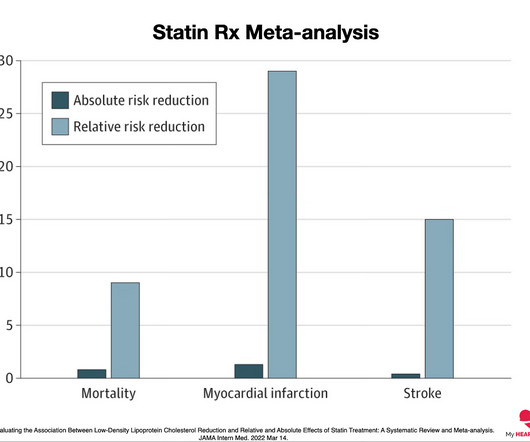
A heart attack is when that plaque ruptures and stops blood flow down the artery. This graph shows the absolute and relative risk reduction for statin therapy in preventing heart attacks (MyocardialInfarction), strokes and preventing death from any cause (All-cause mortality) 2. 2022 Mar 14. J Am Heart Assoc.
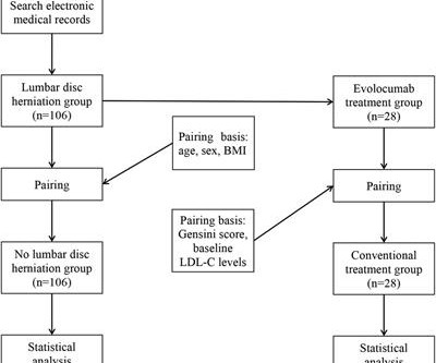
Objective Assessing the impact of lumbar disc herniation (LDH) on the plaque burden of coronary atherosclerosis is our objective. Methods In this study, a total of 212 patients (age 46–80 years) with unstable angina (UA) who underwent coronary angiography (CAG) in our hospital from January 2018 to July 2022 due to UA were included.
2 Trends and Predictors of Participation in Cardiac Rehabilitation Following Acute MyocardialInfarction: Data From the Behavioral Risk Factor Surveillance System. 7 Secondary prevention following myocardialinfarction: a clinical update. 11 Efficacy and Safety of Low-Dose Colchicine after MyocardialInfarction.
Detailed analysis of the excised carotid plaques were carried out with pyrolysis-gas chromatography-mass spectrometry, stable isotope analysis, and electron microscopy. Primary endpoint of the study was a composite of myocardialinfarction, stroke, or death from any cause in those who had micro and nanoplastics in the carotid plaque.
Atherosclerotic cardiovascular disease (ASCVD), caused by plaque buildup in arterial walls, is one of the leading causes of disability and death worldwide.1,2 3 Patients with ASCVD are at a higher risk for major adverse cardiovascular events (MACE) including heart attack or myocardialinfarction (MI), stroke, and cardiovascular (CV) death.4
A CTCA provides much more anatomical detail and can identify advanced plaque often missed by CT Coronary Artery Calcium Score scans alone. There are 3 types of coronary atherosclerosis visible on CTCA: Calcified Plaque - Easily Identified on both CT CAC & CTCA scans. Subscribe now How Often Does A CT CAC Scan Miss Plaque?
While the total body seems to do little in determining cholesterol levels, what is more scientifically shocking is slope of the curve between blood LDL levels and plaque burden is rarely linear. Nat Cardiovasc Res 1 , 554–561 (2022). LDL is obviously a target against atherosclerosis. Mind you LDL constitutes.000025% Reference 1.Lib
Note: the 2022 ACC Expert consensus Chest pain guidelines state that "posterior STEMI-Equivalent" is a sign of acute coronary occlusion. Smith : clearly diagnostic of posterior and high lateral OMI (ST depression V2-V4, with hyperacute T-wave in aVL and recriproal STD in inferior leads).
Influenza-like illness can also trigger plaque rupture. Prevalence and outcome of patients with non-ST segment elevation myocardialinfarction with occluded culprit artery - a systemic review and meta-analysis. This means that, in the United States alone, of 500,000 NSTEMI, 170,000 do not get the care they deserve.
This was attributed to a "Type 2 MI", which is acute MI that is not due to ruptured plaque, but rather due to "supply demand oxygen mismatch". Most MINOCA is due to ruptured plaque with thrombus that lyses and does not leave behind a visible culprit. They made a final diagnosis of type II myocardialinfarction.
The authors describe a case with some features in common with our patient -- a stressful event followed by a stress cardiomyopathy/acute myocardialinfarction overlap syndrome. Acute myocardialinfarction: an uncommon complication of takotsubo cardiomyopathy. Mechanisms of plaque formation and rupture. SanzRuiz, R.,
We organize all of the trending information in your field so you don't have to. Join thousands of users and stay up to date on the latest articles your peers are reading.
You know about us, now we want to get to know you!
Let's personalize your content
Let's get even more personalized
We recognize your account from another site in our network, please click 'Send Email' below to continue with verifying your account and setting a password.


Let's personalize your content