This site uses cookies to improve your experience. To help us insure we adhere to various privacy regulations, please select your country/region of residence. If you do not select a country, we will assume you are from the United States. Select your Cookie Settings or view our Privacy Policy and Terms of Use.
Cookie Settings
Cookies and similar technologies are used on this website for proper function of the website, for tracking performance analytics and for marketing purposes. We and some of our third-party providers may use cookie data for various purposes. Please review the cookie settings below and choose your preference.
Used for the proper function of the website
Used for monitoring website traffic and interactions
Cookie Settings
Cookies and similar technologies are used on this website for proper function of the website, for tracking performance analytics and for marketing purposes. We and some of our third-party providers may use cookie data for various purposes. Please review the cookie settings below and choose your preference.
Strictly Necessary: Used for the proper function of the website
Performance/Analytics: Used for monitoring website traffic and interactions
doi: 10.1136/bmjhci-2022-100718 FDA 510(k) Summary, K233409 Bachtiger, P., for detection of LVEF below 40%, 84.8% sensitivity, and 69.5% specificity when deployed on over 1,050 patients across multiple real-world settings. BMJ Health & Care Informatics, 30:e100718.
The algorithm uses deep learning to analyse routine ultrasound scans of the heart ( echocardiograms ) to detect disease that often goes undetected during standard assessments.
Echocardiogram showed LVEF 66% with normal wall motion and normal diastolic function. The above said — it may prove insightful to take another look at the Wellens' Syndrome case instantly recognized by Dr. Smith in the August 12, 2022 post in Dr. Smith’s ECG Blog. Lesions less than 70% are generally considered to be non-flow limiting.
Hopefully a repeat echocardiogram will be performed outpatient. Retrieved July 2, 2022, from [link] Moyé, D. Retrieved July 2, 2022, from [link] Sybrandy, K. I've copied KEY points from My Comment in the August 6, 2022 post in Dr. Smith's ECG Blog — regarding the answer to this question. No cardiac MRI was done.
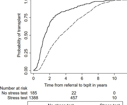
Methods This is a retrospective cohort study of 22 687 end-stage renal disease patients from 2011 to 2022, within an integrated health system. times higher rate of stress echocardiogram (RR 6.51, 95% CI 5.65 Compared with dialysis patients, transplant patients had a 5.6 to 5.92), a 6.5 to 0.58).
It should be treated as such unless there is more information such as old or serial EKGs that can confirm a benign diagnosis, as BTWI patterns can mimic the South Africa Flag Sign (Compare this EKG to case 4 here: [link] com/2022/05/quiz-post-which- of-these-if-any-are-omi.html ).
The LPA to RPA ratio on initial and most recent echocardiogram, intervention on the LPA at initial surgery and subsequent reintervention on the LPA were compared between the two groups.Results:The 22q11 deletion and control group had a similar mean age at time of study 6.9±3.4 0.27, p=0.002) echocardiogram.
We report results from the mavacamten REMS database (28-Apr-2022 to 27-Feb-2024).Methods:Data On the 29,111 status forms in these patients, each representing an assessment of an echocardiogram, LVEF <50% was reported on 276 (0.9%) and HFH was reported on 86 (0.3%). were women; 64.6% were >60 years of age.
We carried out a prospective national specialist nurses in organ donation (SNOD) audit of UK donor offers between 20 August and 31 November 2022, and a prospective national recipient transplant centre audit of all donor offers between 22 September and 19 December 2022. hours (interquartile range [IQR] 13.9–33.2).
So the artery had completely spontaneously reperfused prior to intervention; the duration of occlusion was perhaps 2 hours The troponin peaked at 60,000 ng/L (a very large infarction) Formal bubble contrast echocardiogram --The estimated left ventricular ejection fraction is 46%. Regional wall motion abnormality-inferior.
Results 48 patients (71% with a Fontan circulation, 42% females, mean age 33±9 years) underwent two CPETs between May 2018 and May 2022 with echocardiograms performed within 6 months of each CPET. Apple Watch was the predominant smartwatch used (79%).
Methods In a retrospective cohort study, we used an NLP pipeline applied to the Electronic Health Record (EHR) to identify patients with a clinical diagnosis of HF between 2010-2022. Of 3727 consecutive patients with HF and LVEF ≥ 50% on echocardiogram, only 8.3% Results were validated in a second, independent centre.
Formal Echocardiogram: Normal left ventricular size and wall thickness. As I emphasized in My Comment at the bottom of the page in the October 10, 2022 post in Dr. Smith's ECG Blog — Interpretation of a post-resuscitation ECG can be extremely challenging. First — Some thoughts on the post -resuscitation ECG.
The echocardiogram showed a normal EF without any abnormalities. I suspect there is LA-RA Lead Reversal ( See My Comment in the August 17, 2022 post of Dr. Smith's ECG Blog for review of the effects of LA-RA reversal ). Troponins were all negative. There was no apparent reversible cause found for the worsening heart block.
The patient was thought to have low likelihood of ACS, and cardiology recommended repeat troponin, urine drug testing, and echocardiogram. Bedside echocardiogram showed hypokinesis of the mid to distal anterior wall and apex. Initial hscTnI was 10 ng/L (ref. <14). There was no recommendation for repeat ECG.
The emergent echocardiogram showed normal EF, no WMA, and normal valve function. Unfortunately, this fooled the Emergency Physician and Cardiologist into an emergent angiogram for perceived "inferior STEMI." The angiogram showed completely normal coronary arteries. They rhythm returned to sinus reportedly after metoprolol IV was given.
Echocardiogram: The estimated left ventricular ejection fraction is 34% Regional wall motion abnormality-lateral, akinetic. The February 10, 2022 post in Dr. Smith's ECG Blog — My Comment ( at the bottom of the page ) illustrates the Mirror Test in a case with posterior reperfusion waves ( ie, tall anterior T waves ).
He visited an outpatient clinic for it and an echocardiogram and exercise stress test was normal. In the meantime, cardiology consultant sees the patient and performs a bedside echocardiogram which revealed no major wall motion abnormalities. He has 40 packs-year of smoking history. He denies taking any medication. doi: 10.5543/tkda.2021.21026.
He had his echocardiogram done already and was normal. 2022 Jan 5;11(1):279. I don’t know, whether a single blocked P could by any way a concealed Wenke -Bach. I didn’t have calipers to measure the PR accurately though. The baseline heart rate was around a vago-genic 60/mt, that was comforting. J Clin Med.
Elevated troponins prompted an echocardiogram — which revealed an apical wall motion abnormality (WMA). Patient #1 in today's post did not get expert ECG interpretation. Despite active CP — cath lab activation was deferred and this patient was transported to a local hospital without PCI capability.
Next day echocardiogram showed inferolateral hypokinesia with an EF of %45-50. On echocardiogram you will not see a "posterior" hypokinesia (will see "inferolateral") and, as in this case, LCx may not give the blood supply of basal inferior segment (formerly called "posterior"). 2022 Mar-Apr;71:44-46. Epub 2022 Jan 31.
Formal echocardiogram showed normal EF, no wall motion abnormalities, no pericardial effusion. The patient proceeded to cath where all coronaries were described as normal with no evidence of any CAD, spasm, or any other abnormality. No more troponins were done. He was found to be influenza positive. 1849 after cath: Brugada pattern is gone!
Her contrast enhanced echocardiogram is shown below in the parasternal short axis view. After returning from lab repeat troponin was 20,380 ng/L, and later that evening it peaked at 29,571 ng/L before trending down. The patient suffered a large infarct. Clin Cardiol [Internet].
The echocardiogram shows a preserved left ventricular ejection fraction (LVEF) of 55% with marked basal and mid inferolateral and basal anterolateral hypokinesia. Successful primary angioplasty of the mid-circumflex artery towards the main marginal branch with the implantation of a drug-eluting stent. Good angiographic result.
An echocardiogram was done. Blunt Trauma in a Child 40-something male in a head-on Motor Vehicle Collision and Splenic Injury == MY Comment, by K EN G RAUER, MD ( 10/10 /2022 ): == Highly interesting post by Dr. Smith regarding a 30-something male with multiple injuries from a motor vehicle accident. Is there also Brugada?
The diagnosis was a bit hard to find in the chart, and the echocardiogram did only stated "assymetric hypertrophy." Re ECG recognition of SA Block — See My Comment at the bottom of the page in the May 25, 2022 post — as well as my comment in the Addendum of the August 30, 2023 post.
Formal Echocardiogram: The estimated left ventricular ejection fraction is 58 %. 2022 Jan;51:384-387. A DDENDUM ( 2/8/2022 ): Dr. Mario Parrinello , an esteemed cardiology colleague of ours from Cremona, Italy — wrote the following comment regarding today’s post on the EKG Club. Left ventricular hypertrophy concentric.
Here is the post PCI EKG: And a few hours after that: The post PCI echocardiogram showed: Normal estimated left ventricular ejection fraction, 57%. This is a large OMI that has Zero ST Elevation but can be diagnosed by ECG features other than ST Elevation!! Regional wall motion abnormality-mid to basal inferior wall.
See this case: what do you think the echocardiogram shows in this case? Routine STEMI activation in STE-aVR for emergent revascularization is not warranted, although urgent, rather than emergent, catheterization appears to be important. == MY Comment, by K EN G RAUER, MD ( 11/4 /2022 ): == Our thanks to Drs.
Indeed, bedside Echocardiogram revealed severe left ventricular impairment of Takotsubo cardiomyopathy. Surawicz and Knilans report that intense catecholamine surge, or severe maladjustment of the autonomic nervous system, can manifest “cerebral T waves” in the absence of an acute intracranial process. potassium) were within normal parameter.
This latest data dump did not arrive soon enough to affect the ACIP committee’s recommendations but should serve to chill the ambition of exuberant, overconfident bureaucrats still seeking to mandate vaccines in the fall of 2022. That work is far from complete. Anish Koka is a cardiologist.
While awaiting transfer to the cath lab, STAT echocardiogram was performed and showed LVEF 30-35%, as well as anterior, inferior, and apical hypokinesis, and apical thrombus. The October 21, 2022 post — for " artifactual VT". This confirms the suspicion of prior anterior OMI. The thrombus is circled below in red.
After discussing all of the above with ED staff, we have made a decision to get stat echocardiogram and assess overall LV function and wall motion abnormalities and defer cath lab activation at the time." Precordial Swirl in ECG #1: As discussed in the October 15, 2022 post in Dr. Smith's ECG Blog — Drs. It does not radiate.
Here is the cath report: Echocardiogram: There is severe hypokinesis of entire LV apex and apical segment of all the walls. Hospital Course The patient was taken emergently to the cath lab which did not reveal any significant coronary artery disease, but she was noted to have reduced EF consistent with Takotsubo cardiomyopathy.
She had an echocardiogram which was normal. She was prescribed oral diltiazem to prevent recurrence and was discharged. == MY Comment, by K EN G RAUER, MD ( 10/25 /2022 ): == Today’s blog post reviews the important topic of how to approach the patient who presents with palpitations from an SVT ( S upra V entricular T achycardia ) rhythm.
For instance, the average waiting time for an echocardiogram at Turin’s Molinette Hospital was 31 days in 2016 and an even longer 53 days for a Holter ECG. Prior to the new regulation, getting a consultation with a cardiologist or getting a needed diagnostic cardiology test often involved long waiting times.
Approximately 39 million people were living with HIV at the end of 2022, according to the World Health Organization (WHO). All underwent MRI to measure coronary vessel wall thickness and an echocardiogram to assess left ventricular function. million lives so far.
All these three cases were observed with a severe regurgitation by echocardiogram in the last follow-up. Regular follow-up by echocardiogram is critically important for these patients. ResultsIn our cohort, the female/male ratio was 7:21, with an average age of 8.76.0 0.7526) years.
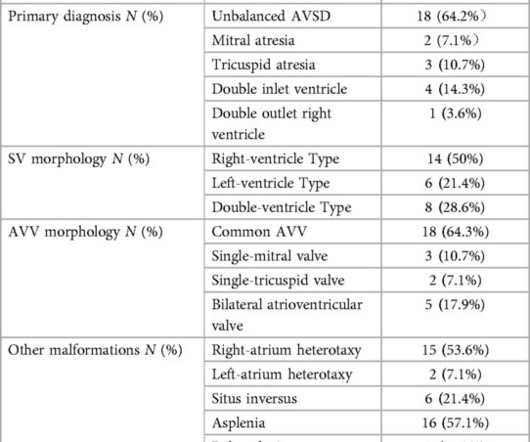
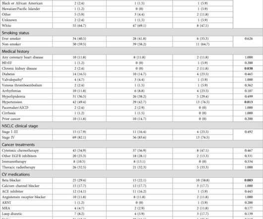
Retrospective analysis of NSCLC patients with 1 echocardiogram post-osimertinib between 2007 and 2022 was performed. However, the nature of cardiac remodeling and associated risk factors remains incompletely understood.
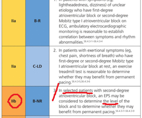
It is reasonable to perform an echocardiogram to evaluate LV function. References: [1] 2022 ESC Guidelines for Ventricular Arrhythmias : Key Points - American College of Cardiology. 2022, September 2) [2] Ward, R. 1 ] Considerations Regarding Use of Flecainide: A 12-lead ECG is mandatory before starting therapy. Van Zyl, M., &
More troponin values were measured at the cardiac center: 2327- 267 ng/L 0821- 355 ng/L 1108- 305 ng/L An echocardiogram on day three of the patients admission showed an ejection fraction of 46% with abnormal basal inferior and basal lateral segments, and severe aortic stenosis.
In this study of consecutive patients with LBBB who were hospitalized and had an echocardiogram, a QRS duration less than 170 ms (n = 262), vs. greater than 170 ms (n = 38), was associated with a significantly better ejection fraction (36% vs. 24%). So indeed the QRS is approximately 200 ms. Comment: What is the normal QRS duration in LBBB?
We organize all of the trending information in your field so you don't have to. Join thousands of users and stay up to date on the latest articles your peers are reading.
You know about us, now we want to get to know you!
Let's personalize your content
Let's get even more personalized
We recognize your account from another site in our network, please click 'Send Email' below to continue with verifying your account and setting a password.


Let's personalize your content