This site uses cookies to improve your experience. To help us insure we adhere to various privacy regulations, please select your country/region of residence. If you do not select a country, we will assume you are from the United States. Select your Cookie Settings or view our Privacy Policy and Terms of Use.
Cookie Settings
Cookies and similar technologies are used on this website for proper function of the website, for tracking performance analytics and for marketing purposes. We and some of our third-party providers may use cookie data for various purposes. Please review the cookie settings below and choose your preference.
Used for the proper function of the website
Used for monitoring website traffic and interactions
Cookie Settings
Cookies and similar technologies are used on this website for proper function of the website, for tracking performance analytics and for marketing purposes. We and some of our third-party providers may use cookie data for various purposes. Please review the cookie settings below and choose your preference.
Strictly Necessary: Used for the proper function of the website
Performance/Analytics: Used for monitoring website traffic and interactions
A 50-something male with hypertension and 20- to 40-year smoking history presented with 1 week of stuttering chestpain that is worse with exertion, which takes many minutes to resolve after resting and never occurs at rest. At times the pain does go to his left neck. It is a ssociated with mild dyspnea on exertion.
Written by Jesse McLaren Four patients presented with chestpain. 2022 ; 51 : 384 - 387 3. 2022 ; 55 : 180 - 182 6. All initial ECGs were labeled ‘normal’ or ‘otherwise normal’ by the computer interpretation, and below are the ECGs with the final cardiology interpretation. Am J Emerg Med. J Electrocardiol. Am J Emerg Med.
Written by Jesse McLaren Two patients in their 70s presented to the ED with chestpain and RBBB. Patient 1 : a 75 year old called paramedics with one day of left shoulder pain which migrated to the central chest, which was worse with deep breaths. Do either, both, or neither have occlusion MI? Vitals were normal.
A 50-something man presented in shock with severe chestpain. Case continued A bedside ultrasound showed diminished LV EF and of course bradycardia. The April 17, 2022 post ( Leads V1,V2 misplacement ). The May 24, 2022 post ( LA-LL reversal ). The May 26, 2022 post ( LA-LL reversal ).
Submitted and written by Anonymous, edits by Meyers and Smith A 50s-year-old patient with no known cardiac history presented at 0045 with three hours of unrelenting central chestpain. The pain was heavy, radiated to her jaw with an associated headache. Triage VS: 135/65 mmHg, 95 bpm, 94% on room air, 16/min, 98.6 Lupu L, et al.
Healthy male under 25 years old with a pretty good story for acute onset crushing chestpain relieved with nitro. No pericardial effusion on ultrasound." PEARL: Most patients who present with new chestpain + ECG changes + positive troponin — will not need Cardiac MRI. What do you think?
They had difficulty describing their symptoms, but complained of severe weakness, nausea, vomiting, headache, and chestpain. They described the chestpain as severe, crushing, and non-radiating. Altogether, this strongly suggests inferolateral OMI, particularly in a patient with acute chestpain.
Submitted and written by Megan Lieb, DO with edits by Bracey, Smith, Meyers, and Grauer A 50-ish year old man with ICD presented to the emergency department with substernal chestpain for 3 hours prior to arrival. At this time he reported ongoing chestpain and was given aspirin and nitroglycerin. J Am Heart Assoc.
Submitted and written by Quinton Nannet, MD, peer reviewed by Meyers, Grauer, Smith A woman in her 70s recently diagnosed with COVID was brought in by EMS after she experienced acute onset sharp midsternal chestpain without radiation or dyspnea. Bedside ultrasound is another very important piece. Do you activate the Cath Lab?
He was given aspirin and sublingual nitro and the pain resolved. Bedside cardiac ultrasound with no obvious wall motion abnormalities. Another ECG was recorded after the nitroglycerine and now without pain: All findings are resolved. He had a previous ECG on file: Proving the findings are new The cath lab was activated.
Most cases go undiagnosed until the condition advances enough to create symptoms such as shortness of breath, chestpain or fatigue. AI algorithms could further improve care by using data from ultrasounds and other imaging devices to develop a digital twin of each patient. “The
Answer : Bedside ultrasound! Smith : RV infarct may also have this appearance on ultrasound. So hypoxia without B lines on lung ultrasound strongly weights toward PE. But the History in today's case was acute shortness of breath with dizziness and lightheadedness — and, essentially without chestpain!
He arrived in the ED and had an immediate bedside cardiac ultrasound while this ECG was being recorded. The bedside ultrasound (video not available) reportedly showed only a slightly reduced LV function. The patient was given 6mg, then 12 mg, of adenosine, without a change in the rhythm. Here is the ECG: What do you think?
If you saw this ECG only knowing that it is an acute chestpain patient, what would be your interpretation? However, in the context of the first ECG and the waning chestpain, this is diagnostic of reperfusion. Due to the severity of the pain and the high BP, they obtained an aortic dissection CT.
Written by Pendell Meyers and Peter Brooks MD A man in his 30s with no known past medical history was reported to suddenly experience chestpain and shortness of breath at home in front of his family. Chestpain, SOB, Precordial T-wave inversions, and positive troponin. Now another, with ultrasound. This is a quiz.
He had concurrent sharp substernal chestpain that resolved, but palpitations continued. Over past 3 months, he has had similar intermittent episodes of sharp chestpain while running, but none at rest. Pads were placed with ultrasound guidance, so they were in the correct position. However, this is not SVT.
24: Joint American College of Cardiology/Journal of the American College of Cardiology Late-Breaking Clinical Trials (Session 402) Saturday, April 6 9:30 – 10:30 a.m.
This case was provided by Spencer Schwartz, an outstanding paramedic at Hennepin EMS who is on Hennepin EMS's specialized "P3" team, a team that receives extra training in advanced procedures such as RSI, thoracostomy, vasopressors, and prehospital ultrasound. On medic arrival, she walked out of the house in no distress, but was diaphoretic.
Written by Willy Frick A 40 year old woman was at home cooking when she developed chestpain. Fortunately, this operator used intravascular ultrasound (IVUS). Unfortunately, a few hours later the patient complained of recurrent chestpain. She took an oxycodone and called EMS. I sent this to Drs.
A 70-something female with no previous cardiac history presented with acute chestpain. She awoke from sleep last night around 4:45 AM (3 hours prior to arrival) with pain that originated in her mid back. She stated the pain was achy/crampy. Over the course of the next hour, this pain turned into a pressure in her chest.
The patient in today’s case is a previously healthy 40-something male who contacted EMS due to acute onset crushing chestpain. The pain was 10/10 in intensity radiating bilaterally to the shoulders and also to the left arm and neck. Written By Magnus Nossen — with edits by Ken Grauer and Smith. The below ECG was recorded.
The best course is to wait until the anatomy is defined by angio, then if proceeding to PCI, add Cangrelor (an IV P2Y12 inhibitor) I sent the ECG and clinical information of a 90-year old with chestpain to Dr. McLaren. His response: “subendocardial ischemia. A emergent cardiology consult can be helpful for equivocal cases.
There was no chestpain. So we did a bedside cardiac ultrasound. I’ve reviewed My Take on the ECG diagnosis of RVH on a number of occasions in Dr. Smith’s ECG Blog ( See My Comment at the bottom of the page in the March 6, 2022 and September 1, 2020 posts , to name just 2 ). But today's patient had no chestpain.
Dr. Nossen performed a bedside ultrasound which was interpreted as normal. See these similar cases: A man in his sixties with chestpain Why is there inferior ST elevation, and would you get posterior leads? Sudden CP and SOB with Inferior ST Elevation and in STE in V1. Is it inferior and RV OMI?
She denied chestpain and denied feeling any palpitations, even during her triage ECG: What do you think? My bedside ultrasound was of insufficient quality, but showed somewhat reduced overall EF, distended IVC without respiratory variation, no pericardial effusion, and diffuse bilateral B lines. == What do you think of her ECG?
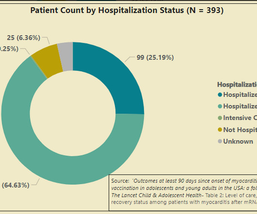
The current study should dispel the ludicrous notion that clinical myocarditis - a disease entity that comes to light when you have chestpain because cells in your heart are dying — is mild. The CDC study published in Lancet on previously healthy 12-29 years old’s is a survey-based study. That work is far from complete.
She did notice something slightly wrong subjectively, but had no palpitations, chestpain, or SOB, or any other symptom. Her bedside cardiac ultrasound was normal We decided to cardiovert her since the time of onset was very recent. 25, 2022 ). Her Apple Watch suddenly told her that she is in atrial fibrillation.
Given her reported chestpain, shortness of breath, and syncope, an ECG was quickly obtained: What do you think? A bedside cardiac ultrasound was performed with a parasternal long axis view demonstrated below: There is a large pericardial effusion with collapse of the right ventricle during systole. She has already had syncope.
Written by Willy Frick A 51 year old man with hypertension presented with three hours of acute onset, severe midsternal chestpain associated with two episodes of nausea and vomiting. or 2) Inferior and lateral OMI that is beginning to reperfuse, even though the patient still has chestpain? ECG 1 What do you think?
A 69 year old woman with a history of hypertension presented to the emergency department by EMS for evaluation of chestpain and shortness of breath. She awoke in the morning with sharp chestpain which worsened throughout the morning. As her pain worsened, so did her dyspnea. This was written by Hans Helseth.
We organize all of the trending information in your field so you don't have to. Join thousands of users and stay up to date on the latest articles your peers are reading.
You know about us, now we want to get to know you!
Let's personalize your content
Let's get even more personalized
We recognize your account from another site in our network, please click 'Send Email' below to continue with verifying your account and setting a password.


Let's personalize your content