This site uses cookies to improve your experience. To help us insure we adhere to various privacy regulations, please select your country/region of residence. If you do not select a country, we will assume you are from the United States. Select your Cookie Settings or view our Privacy Policy and Terms of Use.
Cookie Settings
Cookies and similar technologies are used on this website for proper function of the website, for tracking performance analytics and for marketing purposes. We and some of our third-party providers may use cookie data for various purposes. Please review the cookie settings below and choose your preference.
Used for the proper function of the website
Used for monitoring website traffic and interactions
Cookie Settings
Cookies and similar technologies are used on this website for proper function of the website, for tracking performance analytics and for marketing purposes. We and some of our third-party providers may use cookie data for various purposes. Please review the cookie settings below and choose your preference.
Strictly Necessary: Used for the proper function of the website
Performance/Analytics: Used for monitoring website traffic and interactions
Sent by anonymous, written by Pendell Meyers, reviewed by Smith and Grauer A man in his 40s presented to the ED with HTN, DM, and smoking history for evaluation of acute chestpain. He was eating lunch when he had sudden onset chest pressure, 9/10, radiating to his back, with sweating and numbness in both hands.
Written by Magnus Nossen with Edits by Grauer and Smith The ECGs in today’s case are from 3 different patients all presenting with new-onset CP ( ChestPain ). Elevated troponins prompted an echocardiogram — which revealed an apical wall motion abnormality (WMA). Patient #1 in today's post did not get expert ECG interpretation.
Upon questioning patient, he denies having any chestpain or chest tightness of any sort. In the absence of chestpain and negative troponin , it appears less likely that he is having acute coronary syndrome though EKG appears concerning. Pericarditis would be even more unlikely in someone without chestpain.
He arrived to the ED by helicopter at 1507, about three hours after the start of his chestpain while chopping wood around noon. He arrived to the ED by ambulance at 1529, only a half hour after the start of his chestpain around 1500 while eating. It is also important to recognize that BTWI patterns can be very dynamic.
Formal Echocardiogram: Normal left ventricular size and wall thickness. As I emphasized in My Comment at the bottom of the page in the October 10, 2022 post in Dr. Smith's ECG Blog — Interpretation of a post-resuscitation ECG can be extremely challenging. First — Some thoughts on the post -resuscitation ECG.
Hopefully a repeat echocardiogram will be performed outpatient. ECG of pneumopericardium and probable myocardial contusion shows typical pericarditis Male in 30's, 2 days after Motor Vehicle Collsion, complains of ChestPain and Dyspnea Head On Motor Vehicle Collision. Diagnosing myocardial contusion after blunt chest trauma.
A 60 yo with 2 previous inferior (RCA) STEMIs, stented, called 911 for one hour of chestpain. He had no h/o heart failure. Regional wall motion abnormality-inferior. Regional wall motion abnormality-inferolateral. This means posterior in common terminology) --Normal LV cavity size with moderately increased thickness.
This middle aged male with h/o GERD but also h/o stents presented to the ED with chestpain. The initial troponin I returned at 1500 ng/L and another ECG was recorded as the patient complained of 9/10 chestpain at 10 hours after the first Now the T-wave in III is fully upright, suggesting re-occlusion.
Later, I found old ECGs: 5 month prior in clinic: V5 and V6 look like OMI 9 months prior in clinic with no chest symptoms: V5 and V6 look like OMI 1 year prior in the ED with chestpain: V5 and V6 sure look like a STEMI For this ECG and chestpain in the ED, the Cath lab activated. But the angiogram was clean.
A 40-something woman with diabetes and peripheral vascular disease who frequently needs the ED for chronic pain called 911 for sudden severe chestpain. Echocardiogram: The estimated left ventricular ejection fraction is 34% Regional wall motion abnormality-lateral, akinetic. A massive acute OMI.
Written by Willy Frick A 40 year old woman was at home cooking when she developed chestpain. The patient was thought to have low likelihood of ACS, and cardiology recommended repeat troponin, urine drug testing, and echocardiogram. Bedside echocardiogram showed hypokinesis of the mid to distal anterior wall and apex.
This patient, who is a mid 60s female with a history of hypertension, hyperlipidemia and GERD, called 911 because of chestpain. A mid 60s woman with history of hypertension, hyperlipidemia, and GERD called 911 for chestpain. It is also NOT the clinical scenario of takotsubo (a week of intermittent chestpain).
Case A 39-year-old male without prior medical history presents with chestpain that started 2 hours prior to presentation. He says that the pain intensity was 10/10 at home but now about 4/10. Despite the clinical stability and decreasing pain, this patient needs an immediate angiogram. 2022 Mar-Apr;71:44-46.
A 40 something otherwise healthy man presented with substernal chestpain. Formal Echocardiogram: The estimated left ventricular ejection fraction is 58 %. 2022 Jan;51:384-387. It had occurred once 3 days prior and resolved without any medical visit. What do you think? This ECG is DIAGNOSTIC of acute LAD Occlusion.
The best course is to wait until the anatomy is defined by angio, then if proceeding to PCI, add Cangrelor (an IV P2Y12 inhibitor) I sent the ECG and clinical information of a 90-year old with chestpain to Dr. McLaren. See this case: what do you think the echocardiogram shows in this case?
The pneumothorax was expanded with a chest tube At 17 hours, another ECG was recorded: It is now much less dramatic and has the morphology of Type 2 Brugada The hs troponin I peaked at 6500 ng/L -- this strongly suggests myocardial contusion. An echocardiogram was done. Is there also Brugada? Right ventricular prominence.
She reports that she is now unable to vagal out of her palpitations and is having shortness of breath and dull chestpain. She had an echocardiogram which was normal. She reported a prior history of SVT and has previously performed vagal maneuvers at home with symptom resolution. Her initial EKG is below.
Echocardiogram showed LVEF 66% with normal wall motion and normal diastolic function. He did not remember whether he had experienced any chestpain. Two subsequent troponins were down trending. Within a few days, the patient was extubated and was neurologically intact.
They had difficulty describing their symptoms, but complained of severe weakness, nausea, vomiting, headache, and chestpain. They described the chestpain as severe, crushing, and non-radiating. Altogether, this strongly suggests inferolateral OMI, particularly in a patient with acute chestpain.
See this case: Persistent ChestPain, an Elevated Troponin, and a Normal ECG. This is different from nitroglycerin which produces vasodilation and can improve by pain improving myocardial perfusion. Her contrast enhanced echocardiogram is shown below in the parasternal short axis view. At midnight.
He denied chestpain or shortness of breath. In the clinical context of weakness and fever, without chestpain or shortness of breath, the likelihood of Brugada pattern is obviously much higher. Formal echocardiogram showed normal EF, no wall motion abnormalities, no pericardial effusion.
He reported typical chestpain since 4H AM and arrived at our ED at 10h with ongoing chestpain. The echocardiogram shows a preserved left ventricular ejection fraction (LVEF) of 55% with marked basal and mid inferolateral and basal anterolateral hypokinesia. The first ECG (10h14) showed TWI in inferior leads."
His medical history is unremarkable except a similar pain occurred 4-5 times in the previous 3 months with less intensity, short duration, unrelated to exertion. He visited an outpatient clinic for it and an echocardiogram and exercise stress test was normal. He has 40 packs-year of smoking history. He denies taking any medication.
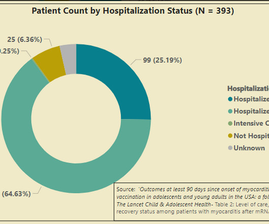
The current study should dispel the ludicrous notion that clinical myocarditis - a disease entity that comes to light when you have chestpain because cells in your heart are dying — is mild. The CDC study published in Lancet on previously healthy 12-29 years old’s is a survey-based study. That work is far from complete.
A 69 year old woman with a history of hypertension presented to the emergency department by EMS for evaluation of chestpain and shortness of breath. She awoke in the morning with sharp chestpain which worsened throughout the morning. As her pain worsened, so did her dyspnea. This was written by Hans Helseth.
He denied chestpain. In this study of consecutive patients with LBBB who were hospitalized and had an echocardiogram, a QRS duration less than 170 ms (n = 262), vs. greater than 170 ms (n = 38), was associated with a significantly better ejection fraction (36% vs. 24%). So indeed the QRS is approximately 200 ms.
We organize all of the trending information in your field so you don't have to. Join thousands of users and stay up to date on the latest articles your peers are reading.
You know about us, now we want to get to know you!
Let's personalize your content
Let's get even more personalized
We recognize your account from another site in our network, please click 'Send Email' below to continue with verifying your account and setting a password.


Let's personalize your content