This site uses cookies to improve your experience. To help us insure we adhere to various privacy regulations, please select your country/region of residence. If you do not select a country, we will assume you are from the United States. Select your Cookie Settings or view our Privacy Policy and Terms of Use.
Cookie Settings
Cookies and similar technologies are used on this website for proper function of the website, for tracking performance analytics and for marketing purposes. We and some of our third-party providers may use cookie data for various purposes. Please review the cookie settings below and choose your preference.
Used for the proper function of the website
Used for monitoring website traffic and interactions
Cookie Settings
Cookies and similar technologies are used on this website for proper function of the website, for tracking performance analytics and for marketing purposes. We and some of our third-party providers may use cookie data for various purposes. Please review the cookie settings below and choose your preference.
Strictly Necessary: Used for the proper function of the website
Performance/Analytics: Used for monitoring website traffic and interactions
Pulmonary hypertension (PH) is a complex and progressive disorder characterised by elevated pulmonary artery pressure. Transcatheter aortic valve implantation (TAVI) is a minimally invasive surgical procedure that has revolutionised the treatment of severe aortic stenosis (AS).
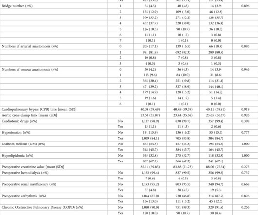
Objective The initial operation for type A aortic dissection has limitations, and there may be a need for reoperation in cases such as giant pseudoaneurysm formation and reduced blood supply to the distal vessels. In this study, we retrospectively analyzed the recorded data of 62 patients.
Patients with significant pulmonary oedema or aortic valve (AV) closure during venoarterial extracorporeal membrane oxygenation (VA-ECMO) were randomized to early left ventricular (LV) unloading or conventional strategy groups (1:1). The primary endpoint was the rate of weaning from VA-ECMO during index admission. vs. 1.7 ± 0.6
Animal studies have shown that mice with TBX1 gene mutations have smaller left pulmonary arteries compared to wild type mice, defined by a reduced left pulmonary artery (LPA) to right pulmonary artery (RPA) ratio. A single study has shown this translates to humans with 22q11 and structurally normal hearts.
33 preoperative clinical features and 4 postoperative complications were collected in each group. LASSO is a regression analysis method that performs both variable selection and regularization to enhance model prediction accuracy and interpretability.
Retrieved July 2, 2022, from [link] Moyé, D. Retrieved July 2, 2022, from [link] Sybrandy, K. Retrieved July 2, 2022, from [link] == MY Comment , by K EN G RAUER, MD ( 2/4 /2024 ): == Today's case by Dr. Small-but-present initial Q waves are seen in 4/6 chest leads ( ie, in leads V2,V3,V4,V5 — as per the RED arrows in Figure-1 ).
Written by Pendell Meyers A woman in her 20s with connective tissue disorder and history of aortic root and valve repair presented with palpitations. Further history revealed she had new onset atrial flutter soon after her aortic surgery, and was put on flecainide approximately 1 month ago. Here is her triage ECG: What do you think?
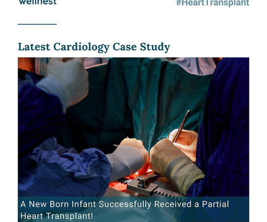
Unlike traditional approaches relying on non-viable cadaver grafts, this procedure involved the transplantation of a portion of the heart, specifically containing the aorta and pulmonary valves, sourced from an infant donor upon cardiac death. Finally, the pulmonary root was transplanted, completing the intricate procedure.
The pipeline of algorithms likely to clear regulatory hurdles and enter the cardiac market over the next 12-18 months include those for Pulmonary Hypertension, Cardiac Amyloidosis, Diastolic dysfunction, and Hyperkalaemia. In April 2022 BMS had received FDA approval for Camzyos, the first drug developed specifically for targeting HCM.
Stenotic lesions included 16 branch pulmonary arteries, 9 aortic isthmus, 2 right ventricular outflow tracts, and 1 Glenn anastomosis. Stent performance was assessed.ResultsWe identified 28 patients (67.8% male) with median age and weight of 3.4 years (interquartile range [IQR], 1.55.5) kg (IQR, 9.116.4).
Event 2023 STS Coding Workshop kchalko Tue, 11/15/2022 - 16:12 Event dates Feb 10–11, 2023 Location Virtual Registrants : To access the library of on-demand content, log into the STS Learning Center. Then go to “In Progress” courses on the dashboard or look in the “My Activities” tab. hours total) Joseph Turek, MD, Jeffrey P.
No signs for aortic dissection or pulmonary embolus. --"Results were discussed with the ordering physician. If there are T-wave inversions and elevated trops in the context of persistent pain, think of other pathologies such as pulmonary embolism. A CT Coronary angiogram was ordered. LAD plaque with 0-25 percent stenosis.
On his physical examination, cardiac and pulmonary auscultation was completely normal. As his pain was very severe, emergency physicians concerned of aortic dissection and ordered a thoracic CT scan. Bi-phasic scan showed no dissection or pulmonary embolism. He has 40 packs-year of smoking history. Turk Kardiyol Dern Ars.
Program Designations Access and Publications (A&P) 1 Participant User File (PUF) 2 Task Force on Funded Research (TFR) 3 Special Projects 4 Adult Cardiac Surgery Database Lead Author Title Publication Date William Keeling 2 National Trends in Emergency Coronary Artery Bypass Grafting European Journal of Cardiothoracic Surgery October 2023 Jake (..)
I suspect pulmonary edema, but we are not given information on presence of B-lines on bedside ultrasound, or CXR findings. Anything that causes pulmonary edema: poor LV function, fluid overload, previous heart failure (HFrEF or HFpEF), valvular disease. What "initiates" the aortic stenosis cascade? She was started on lasix.
His initial high sensitivity troponin I returned at 1300 ng/L and given that his cardiac workup was otherwise unremarkable, a CT was obtained to evaluate for pulmonary embolism and aortic aneurysm or dissection but this too was unrevealing. Also: electrical instability, pulmonary edema, or hypotension.
Larger shunt volume means less blood exiting the left ventricle through the aortic valve and lower cardiac output. Rupture can be either free wall rupture (causing tamponade) or septal rupture, causing ventricular septal defect with left to right flow and resulting pulmonary edema and shock. Cardiology , 114. link] [5] Vega, J.
No thoracic aortic hematoma, aneurysm or dissection. No pulmonary embolism is identified. 2022;Available from: [link] Click here to sign up for Queen of Hearts Access. == MY Comment, by K EN G RAUER, MD ( 10/27/2024 ): == I view today's case as unfortunate. CT Angio Chest IMPRESSION 1.
We organize all of the trending information in your field so you don't have to. Join thousands of users and stay up to date on the latest articles your peers are reading.
You know about us, now we want to get to know you!
Let's personalize your content
Let's get even more personalized
We recognize your account from another site in our network, please click 'Send Email' below to continue with verifying your account and setting a password.


Let's personalize your content